
Presentation
Poor visual acuity with bilateral optic atrophy on the ophthalmological exam.
Patient Data
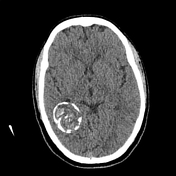

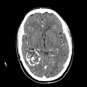
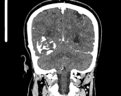
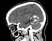

There is a well-circumscribed calcified mass located in the right posterior temporal lobe with arch-like central calcification. No enhancement or surrounding edema is seen. A mass effect is noted on the right atrium with dilated ipsilateral temporal horn.
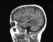
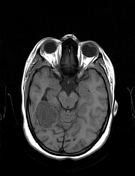
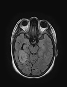
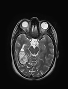

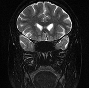
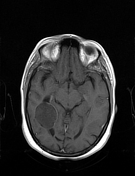
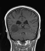
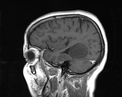
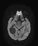
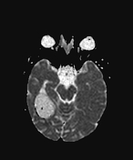
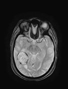

The previously described mass appears of low signal intensity on T1WI, high signal intensity on FLAIR and T2WI with characteristic serpentine linear appearance “snake sign” of the calcified membranes within the mass. The peripheral calcification of the pericyst appears of high signal intensity on T1WI, and low signal intensity on FLAIR, T2WI and gradient echo. No peripheral enhancement or surrounding edema is seen.
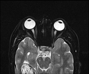
Both optic nerves, as well as the optic chiasma, are atrophied; well-visualized on axial and coronal T2 fat sat.
Case Discussion
With such CT, and MRI features in patient from an endemic echinococcosis area a cerebral calcified hydatid cyst should be suspected.
The differential diagnosis should includes 1:
- calcified brain tumors as meningiomas: The attachement , the dura matter, the perilesional edema, and contrast enhancement seen with tumors are usually absent in hydatid cysts.
- infectious processes such as tuberculosis and bacterial abscess.
- Calcified AVM
The hydatid disease of the central nervous system is seen in around 1% of cases and usually diagnosed during childhood 2. It most frequently located in both hemispheres mainly in the middle cerebral artery territory 2. In calcified hydatid cysts the serological tests are frequently negative 2.


 Unable to process the form. Check for errors and try again.