
Presentation
Macrocephaly and seizures
Patient Data
Age: 7 years
Gender: Female
From the case:
Canavan disease
Show annotations
Download
Info

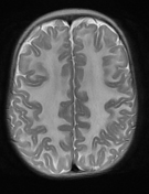
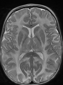
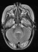
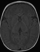
Diffuse T2 hyperintensity at the subcortical U fibers, the bilateral thalami, and the cerebellar peduncles, with sparing of the corpus callosum, caudate nucleus, putamen and internal capsule.
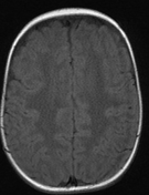
Corresponding T1 hypointensity involving the subcortical U fibers and bilateral thalami.
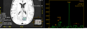
MRS shows elevated NAA peak.
Case Discussion
This case depicts the typical imaging characteristics of Canavan disease.
Note the diffuse T2 hyperintensity involving the white matter at the subcortical U fibers, the bilateral thalami and cerebellar peduncles with corresponding T1 hypointensity.
MRS reveals elevated NAA peak typical for Canavan disease.


 Unable to process the form. Check for errors and try again.