
Presentation
Patient with a history of diabetes mellitus and chronic kidney failure presented with a complain of lumbar pain for several days.
Patient Data
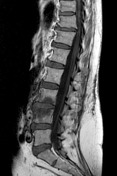

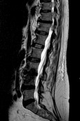
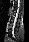
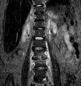
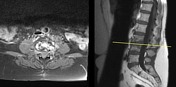

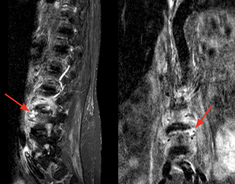
There is a L3-L4 inflammatory process involving the vertebral end plates and bodies. The lesion is hypointense on T1WI, hyperintense on T2WI and STIR and shows an abnormal enhancing. Small paraspinal abscesses are also identified.
Case Discussion
MRI of the lumbar spine was performed and diagnosed spodylitis. The symptoms didn't show any improvement, even after broad-spectrum antibiotic therapy. The patient underwent biopsy and the culture of the material, wich was positive to Candida albicans. Usually, the differentiation between the aetiological agents of spondylitis by MRI is not possible. Lee et al analysed patients with infectious spondylitis who underwent MRI and biopsy from 1998 to 2011. The authors concluded that Candida spondylitis should be considered when infectious lesions contain low-signal spinal inflammatory masses on T2-weighted imaging, small paraspinal abscesses, specially in immunocompromised patients


 Unable to process the form. Check for errors and try again.