
Presentation
MVA. Post laparotomy which was negative
Patient Data


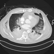
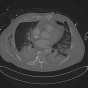

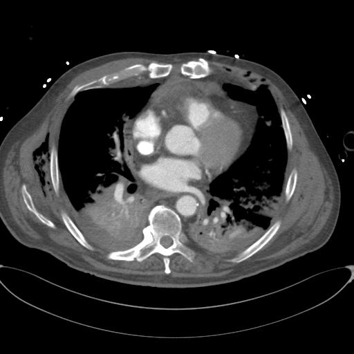
Chest
- ETT. Bilateral ICCs – the left ICC is kinked.
- Small bilateral haemopneumothoraces. The left directly communicates with the chest wall through a displaced fracture of the 5th rib.
- Sternotomy wires are ununited (chronic).
- Traumatic right ventricular outflow tract/pulmonary trunk injury with pseudoaneurysm formation and anterior mediastinal haematoma.
- Small volume anterior pericardial haematoma.
- Adjacent displaced sternal fracture with chest wall haematoma.
- Bilateral lower lobe consolidation.
- Right middle lobe and the lingular contusions.
- Left sided 3rd-11th rib fractures, with flail segments involving the 3rd and 4th ribs.
- Right sided 4th-9th rib fractures. The 7th and 8th are comminuted. There are also undisplaced right 11th and 12th fractures.
- Right crus haematoma - diaphragmatic rupture suspected.
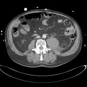
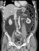
Abdomen/Pelvis
- Gas post laparotomy.
- IDC and left femoral arterial line.
- Small fluid collection surrounds the lower pole of the left kidney with associated perinephric stranding and thickening of the posterior, and to a lesser extent the anterior, pararenal fascia. This most likely represents a ruptured left renal cyst.
- Left psoas haematoma and minor retroperitoneal haematoma - no pancreatic/duodenal injury noted.
- Periportal oedema is likely related to the aggressive fluid resuscitation.
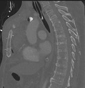
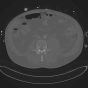
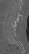
Thoracolumbar spine
- L1 Chance fracture with retropulsion of the inferior fragment resulting in at least 50% spinal canal narrowing. The fracture extends into the superior articular processes bilaterally. L1 spinous process and bilateral transverse process fractures.
- Dislocation of the T12/L1 facet joints bilaterally.
- Fracture of the left T12 inferior articular process.
- L2 superior endplate compression fracture and right L2 transverse process fracture.
Case Discussion
Cardiac injury is one of the most lethal injuries in thoracic trauma. Any of the structures around the heart can be injured. Thus injuries include: traumatic pericardial effusions, pericardial lacerations with or without cardiac luxation (herniation), myocardial transmural or papillary muscle rupture, valvular dysfunction and coronary artery injury 1. Any pericardial effusion detected in the trauma setting must be assumed to be haemopericardium until proved otherwise 1.
In penetrating wounds to the mediastinum, the heart, great vessels, trachea, oesophagus and bony cage of the thorax can all be injured concomitantly. The right ventricle is most commonly injured due to its anterior position in the mediastinum followed by the left ventricle, right atrium then left atrium in penetrating injuries 2.
CT findings of cardiac injury include: haemopericardium, pneumopericardium, pericardial or myocardial lacerations, contrast extravasation into the pericardial sac or mediastinum and displacement of the heart due to cardiac herniation 3.
When a Chance fracture is identified, the abdominal viscera must be carefully inspected as associated intestinal and solid organ injuries are common, especially upper retroperitoneal injuries involving the pancreas and duodenum.


 Unable to process the form. Check for errors and try again.