
Presentation
Acute dyspnoea
Patient Data
Age: 60 years
Gender: Male
From the case:
Cardiogenic pulmonary oedema


Download
Info
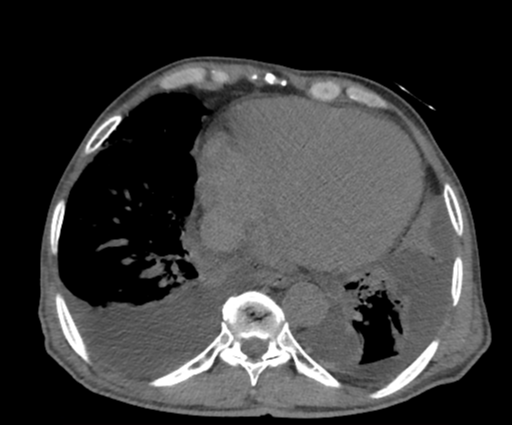
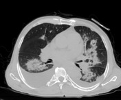
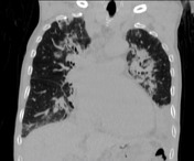
Bilateral airspace opacification in a central peribronchovascular distribution associated with smooth Interlobular septal thickening (which indicates interstitial oedema) and moderate bilateral pleural effusion. Mild cardiac enlargement also noted.
Case Discussion
CT findings are in keeping with acute pulmonary oedema.
The main imaging differential considerations include other causes of diffuse airspace opacification:
- Diffuse pulmonary haemorrhage: has no dependent gradient and usually no pleural effusion
- Pneumonia: usually no dependent gradient
- Pulmonary alveolar proteinosis: usually no pleural effusion


 Unable to process the form. Check for errors and try again.