
Carotid body tumor with lung metastases
Updates to Study Attributes
Pending.
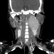
Image CT (C+ delayed) ( update )

Image CT (C+ delayed) ( update )
Updates to Study Attributes
Pending.
Image CT (lung window) ( update )

Image 1 CT (lung window) ( update )
Updates to Study Attributes
Pending.
Image X-ray (Frontal) ( update )

Updates to Study Attributes
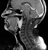
MRI through the neck demonstrates a large mass on the left splaying the ICA and ECA. It is of high T2 signal with numerous punctate and serpiginous regions of low signal due to flow voids (so called salt and pepper sign).
The post contrast T1 fat suppressed images demonstrate vivid enhancement again with flow voids.
Image MRI (T1) ( update )
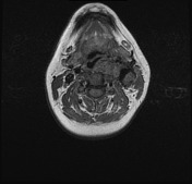
Image MRI (T1 C+ fat sat) ( update )
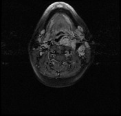
Image MRI (T1) ( update )
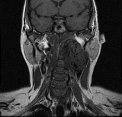
Image MRI (T1 C+ fat sat) ( update )
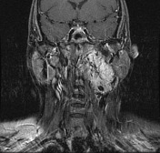
Image MRI (T1) ( update )
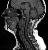
Image MRI (T1 C+ fat sat) ( update )
Updates to Case Attributes
MRI through the neck demonstrates a large mass on the left splaying the ICA and ECA. It is of high T2 signal with numerous punctate and serpignous regions of low signal due to flow voids (so called salt and pepper sign).
The post contrast T1 fat suppressed images demonstrate vivid enhancement again with flow voids.
Features are consistent with a carotid body tumour, in this case metastasising to the lungs.
-<p>MRI through the neck demonstrates a large mass on the left splaying the ICA and ECA. It is of high T2 signal with numerous punctate and serpignous regions of low signal due to flow voids (so called <a href="/articles/salt-and-pepper-sign" title="Salt and pepper sign">salt and pepper sign</a>). </p><p>The post contrast T1 fat suppressed images demonstrate vivid enhancement again with flow voids. </p><p>Features are consistent with a <a href="/articles/carotid-body-tumour" title="Carotid body tumour">carotid body tumour</a>, in this case metastasising to the lungs.</p>- +<p>Features are consistent with a <a href="/articles/carotid-body-tumour">carotid body tumour</a>, in this case metastasising to the lungs.</p>
Tags changed:
- findings


 Unable to process the form. Check for errors and try again.