
Presentation
Right lower abdominal pain, fever and vomiting for 5 days.
Patient Data
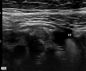
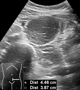
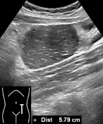
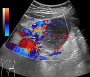
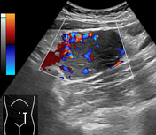

Minimal free fluid in the right iliac fossa. Appendix not confidently identified. Well defined homogeneous hypoechoic mass measuring about 4.5 x 3.9 x 5.8 cm seen in the left lower abdomen, adjacent to the iliac vessels. Color Doppler ultrasound examination shows significant internal vascularity.


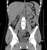
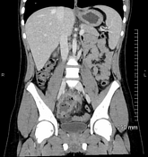
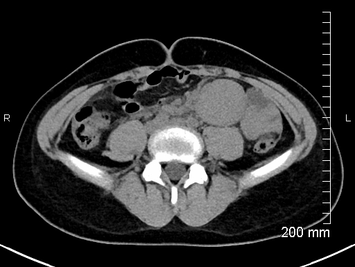
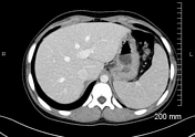
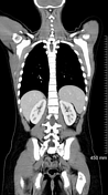
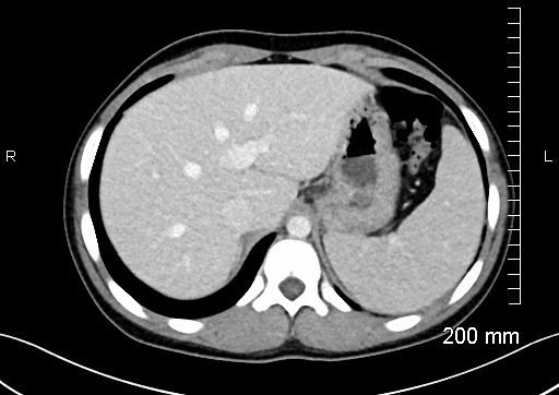
Appendix could not be traced. An irregular collection containing fluid, air densities and a linear opacity (appendicolith?) is noted in the right iliac fossa. Mild free fluid in the pelvis and around the liver. Enhancing soft tissue mass lesion measuring 4.5 x 4.5 x 6.0 cm noted in the root of the mesentery. Prominent veins are seen associated with this lesion which are draining into the adjacent superior mesenteric vein. Multiple enlarged lymph nodes noted along the superior mesenteric vessels.
Mesenteric mass seen in the left lower abdomen on previous study had been excised. Appendicular abscess had been surgically drained. Multiple small subcentimeter cervical and mesenteric lymph nodes; otherwise, no significant lymphadenopathy is seen. Type II retroaortic left renal vein draining in to the IVC at L3 level.
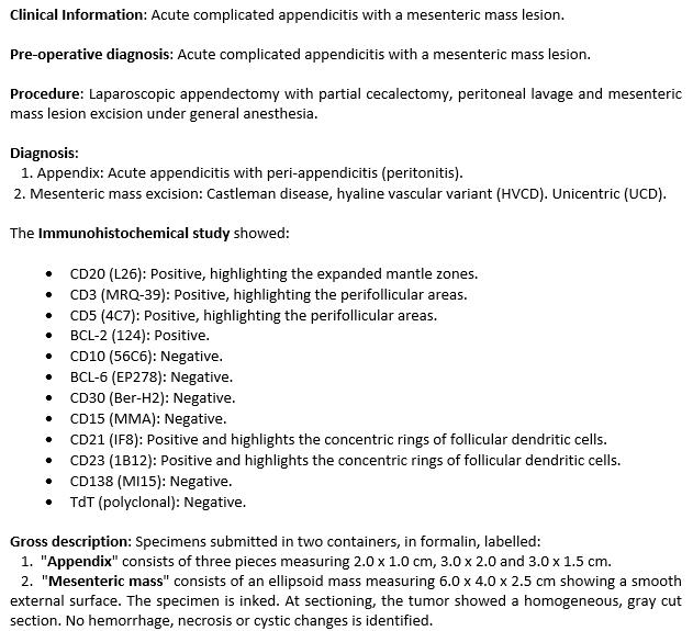
Histopathology report of the excised mesenteric mass lesion and appendectomy specimen.
Case Discussion
Baseline CT scan features are suggestive of ruptured appendix with appendicular abscess formation, which were confirmed intraoperatively. The patient underwent an uneventful appendectomy, draining of the appendicular abscess and excision of the mesenteric mass lesion. After the histopathological diagnosis of the unicentric Castleman disease, the patient was referred to the oncologist for further evaluation.


 Unable to process the form. Check for errors and try again.