
Presentation
The patient presented with a severe headache.
Patient Data
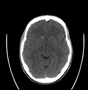

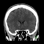
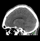

Focal hyper-density of the head of the right caudate nucleus. Small retro-cerebellar arachnoid cyst.
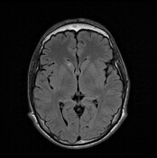
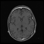
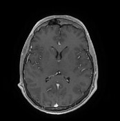
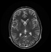
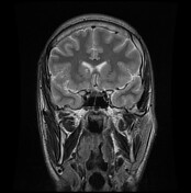
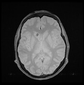
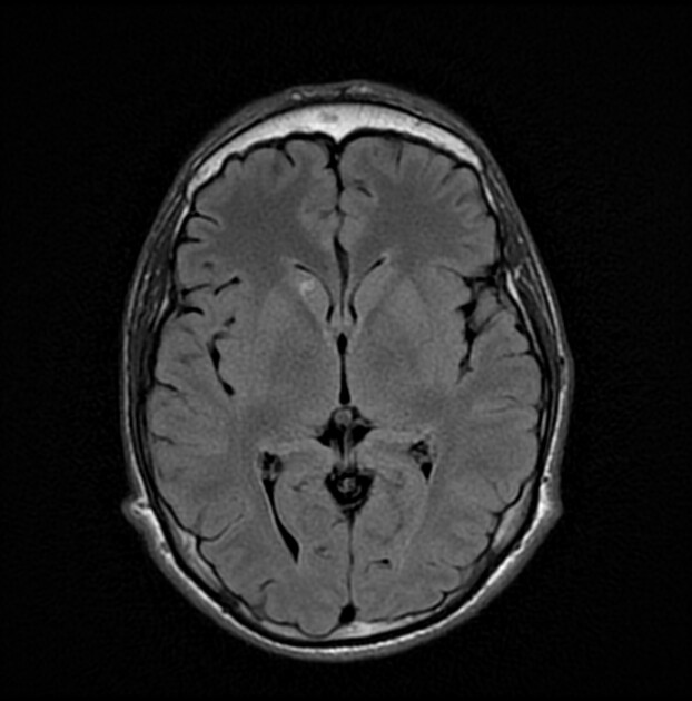
A small ill-defined area of abnormal signal focal area is seen within the head of right caudate nucleus. It exhibits hyper-intense signal at T1, T2 WI, and shows area of blooming at the gradient images denoting blood degradation product. It showed no perifocal edema. No diffusion restriction at DWI.
A well-defined CSF focal area is seen at the medial aspect of right temporal lobe, suggesting prominent Virchow Robin space, rather than old infarct.
Case Discussion
Cavernomas are common cerebral vascular malformations. They are also known as cavernous hemangiomas. Cavernoma is the third most common cerebral vascular malformation after developmental venous anomaly and capillary telangiectasia.


 Unable to process the form. Check for errors and try again.