
Presentation
Hearing loss and was referred to rule out acoustic neuroma.
Patient Data


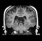
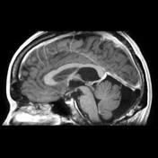
A cystic space is seen in the region of the pineal gland, displacing the fornices upwards and the internal cerebral veins downwards. The pineal gland can be seen as separate from this region, below the internal cerebral veins - best seen on sagittal images. Features are characteristic of a cavum velum interpositum cyst.
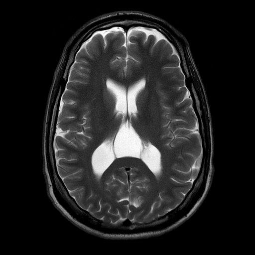
On axial imaging, it is triangular in shape. Incidental note is also made of a large cerebellomedullary cistern (mega cisterna magna).

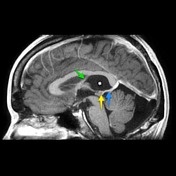
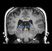
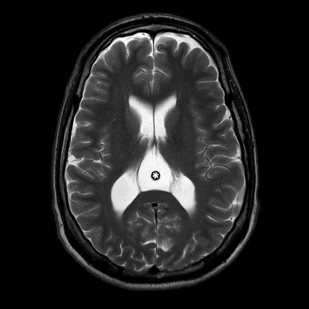
The cavum velum interpositum ( * ) is a triangular space in axial section located below the fornices (green) and above the internal cerebral veins (blue) and thus also above the pineal gland (yellow).
Case Discussion
This case illustrates the typical appearances of a large (cystic) cavum velum interpositum the importance of which is mainly in that it is sometimes confused with pineal regions cysts / cystic tumours. It is often also inappropriately called a cavum vergae, which it is not, as the cavum vergae is located above the fornices.


 Unable to process the form. Check for errors and try again.