
Presentation
Symptoms of increased intracranial pressure. A CT scan was performed (not shown) and a VP shunt was inserted. An MRI was requested two months later.
Patient Data
Age: 40 years
Gender: Male
From the case:
Central neurocytoma


Download
Info
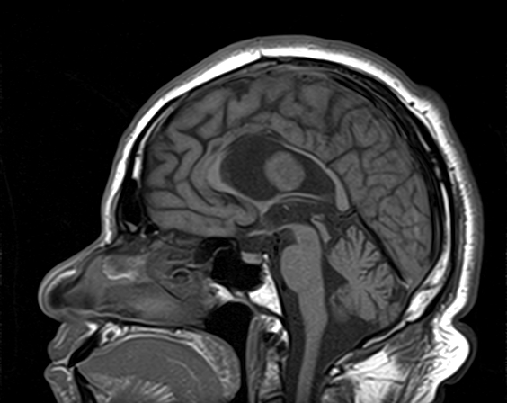
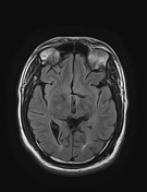
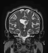
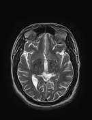
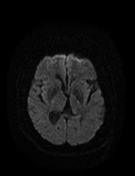
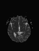
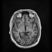
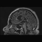
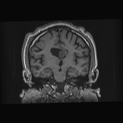
There is a well-defined ovoid soft tissue mass within a dilated right lateral ventricle, attached to the septum pellucidum. It demonstrates a low signal intensity on T1WI, iso-to slightly high signal intensity on T2WI/FLAIR with no enhancement on postcontrast sequences. The adjacent portion of the septum pellucidum appears thickened with same signal intensity. Evidence of restricted diffusion on DWI/ADC.
Case Discussion
MRI features of an intraventricular tumor attached to the septum pellucidum, probably central neurocytoma.


 Unable to process the form. Check for errors and try again.