
Presentation
Persistent headaches.
Patient Data
Age: 20 years
Gender: Female
From the case:
Central neurocytoma


Download
Info
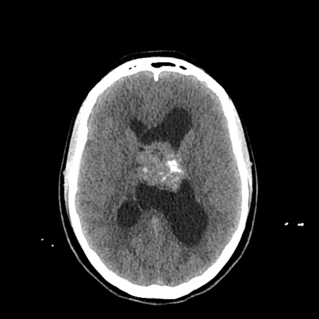
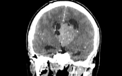
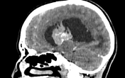
There is a lobulated intraventricular mass expanding the body of the left lateral ventricle extending to the 3rd ventricle through the ipsilateral foramen of Monro. It appears isodense to the cortical grey matter, containing foci of calcification as well as cystic areas with heterogeneous enhancement on postcontrast images.
Case Discussion
CT features are suggestive of a central neurocytoma.
Additional contributor; R. Bouguelaa, MD


 Unable to process the form. Check for errors and try again.