
Presentation
Headache and right side hemiparesis.
Patient Data
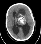

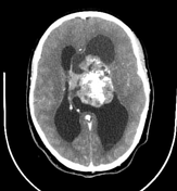
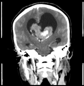

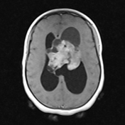
Pre and post contrast brain CT shows a large, heterogeneously hyperdense mass centred in the body of the left lateral ventricle and attached to the septum pellucidum. There are multiple, non-enhancing cystic components and coarse central calcifications. The mass enhances intensely on post contrast images. It has caused marked obstructive hydrocephalus.
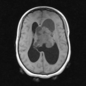
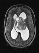
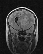

Brain MRI of the same patient demonstrates a heterogenous intraventricular mass in the body of the left lateral ventricle with multiple, small, T2 bright cystic foci giving the mass a bubbly appearance. The cystic areas suppress incompletely on FLAIR. Strong, heterogeneous enhancement of the mass is noted on the post-gadolinum T1 images.
Case Discussion
A calcified, intensely enhancing, intraventricular mass in the body of the lateral ventricle with close attachment to the septum pellucidum in a young patient is fairly characteristic of a central neurocytoma.


 Unable to process the form. Check for errors and try again.