
Presentation
Headaches. Incidental finding.
Patient Data
Age: 65 years
Gender: Female
From the case:
Cerebellar cavernoma


Download
Info

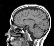
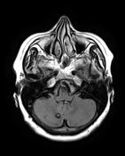
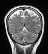
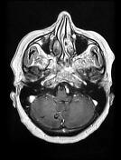
Small rounded well-circumscribed right cerebellar lesion of mixed signal intensity centrally on both T1, T2 and FLAIR with signal loss and blooming on GE sequence. No surrounding edema or enhancement on the postcontrast sequence.
Moderate cerebral volume loss with small vessel ischemic change.
Case Discussion
MRI features characteristic of a cerebellar cavernoma (or cavernous venous malformation), type II according to Zabramski classification.


 Unable to process the form. Check for errors and try again.