
Presentation
The patient presented with ataxia and hearing loss in the left ear after an episode of dizziness while swimming, which led to difficulty moving and needing to drag themselves to the edge of the pool.
Patient Data
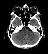


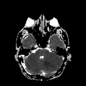
Initial findings at NECT (non-enhanced computed tomography) were cerebellar bilateral focal ischemic hypodense lesions.
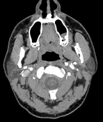
CT angiography showed bilateral areas of alternating narrowing and dilatation in the third segment of both vertebral arteries associated with left vertebral artery ectasia-dominance and left posterior inferior cerebellar (PICA) artery occlusion.
No abnormality was found in the carotid arteries and anterior cerebral artery segments.
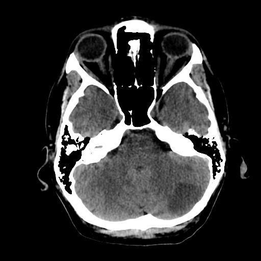
Consequently, NECT (non-enhanced computed tomography) showed subacute left cerebellar hemisphere ischemic infarction.
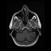
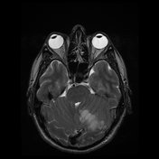
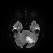
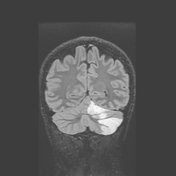
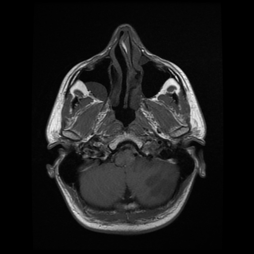
Magnetic resonance imaging showed an ischemic lesion that was hyperintense in T2W images, hypointense in T1W images, hyperintense in DWI images, and hypointense in ADC images.
This was a subacute left cerebellar hemisphere infarction with mass effect and partial compression of the fourth ventricle.
Case Discussion
The vessel occlusion was restored by intravenous thrombolysis with a good clinical recovery.
Stroke in young adults could be caused by vertebral artery fibromuscular dysplasia (FMD), which can affect layers of both small and medium caliber arteries, most frequently the cervical carotid and vertebral arteries, and renal arteries 1. FMD consists of segmental dilatation and focal arterial stenosis and could be complicated or not by dissection, subarachnoid hemorrhage, or arterial occlusion 1.
Vertebral artery dominance (VAD), which is a condition derived from the asymmetry of the vertebral artery diameters on both sides, is a risk factor for posterior circulation ischemic stroke (PCIS) because the association with posterior inferior cerebellar artery infarction is high 2.
Medical management was an essential treatment in this case, however, the natural history of the disease is unpredictable and it is not clear if the arteriopathy will progress or not 3.


 Unable to process the form. Check for errors and try again.