
Presentation
Headache.
Patient Data
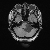

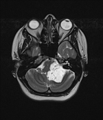
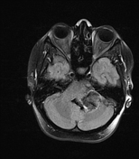
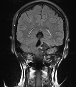
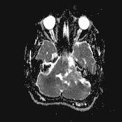
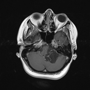
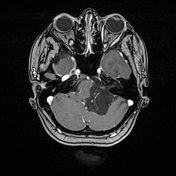
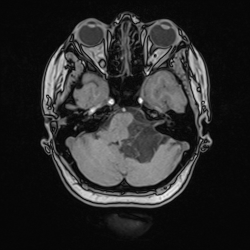
Extra-axial lobulated mass lesion was seen centered at/expanding the left cerebellopontine angle, it insinuates through the left cerebellopontine, pre-pontine, and cerebellomedullary cisterns extending down to the level of the foramen magnum. It encases the left PICA and AICA, intimately related to the intracranial segment of the left vertebral artery, with no frank vertebral/basilar artery encasement, no feature of definite internal auditory canal extension, however, the cisternal segment of the left facial, and vestibulocochlear nerves are encased.
The lesion is heterogenous, shows high SI on T2, of slightly higher signal than CSF on T1, partially attenuated on FLAIR with diffusion restriction slightly less than brain parenchyma (mean ADC of 0.96 × 10 -3mm2 s-1), no solid enhancement post-contrast.
It exerts a noticeable pressure effect manifested as:
deformation of the left cerebellar hemisphere, and middle/inferior cerebellar peduncles
compression of the medulla oblongata with 8-9 mm right-sided midline shift almost effacing the right cerebellomedullary cistern. No intrinsic signal abnormality, no tonsillar herniation
obliterating the left lateral aperture of Luschka with noticeable fourth ventricle prominence, no intra-ventricular extension, and no supratentorial hydrocephalus
Case Discussion
Imaging findings of a left cerebellopontine angle epidermoid cyst.


 Unable to process the form. Check for errors and try again.