
Presentation
6-month history of headaches and right-sided hearing loss.
Patient Data


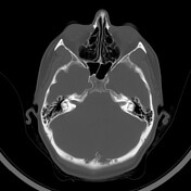
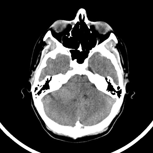
Non-contrast CT scan of the head demonstrates an isodense extra-axial lesion in the right posterior fossa abutting the petrous temporal bone. There is mass effect upon the brainstem with leftward deviation. No associated widening of the internal auditory canal was identified on the right side. There is no hyperostosis of the right petrous temporal bone.
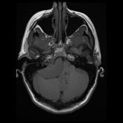
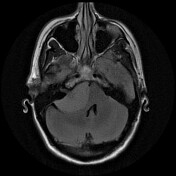

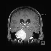
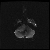
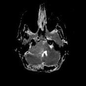
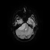

MRI of the brain demonstrates a large enhancing lesion in the right cerebellopontine angle. It is extra-axial with internal auditory canal extension. It has a small dural tail. It is adherent to the right sigmoid-transverse sinus with some compression and no obvious invasion.
Histopathology report
MACROSCOPIC Right cerebellopontine angle tumor: Multiple irregular pieces of tan-brown tissue measuring 25 x 20 x 12 mm in aggregate. The specimen is submitted in toto in 1A to 1C.
MICROSCOPIC Right cerebellopontine angle tumor: The sections show a meningioma of no special type with low grade cytoarchitectural features. The histological features are somewhat compromised by cautery artefact. No cerebral cortex is incorporated in the biopsy.
Case Discussion
The preoperative radiological findings were suspected to represent vestibular schwannoma with a differential of meningioma. The patient underwent craniotomy surgery with histopathology results confirming meningioma.


 Unable to process the form. Check for errors and try again.