
Presentation
Headache
Patient Data


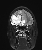
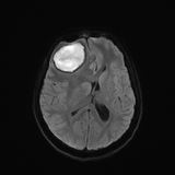
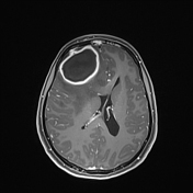
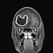
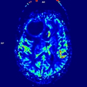
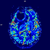
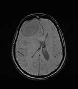
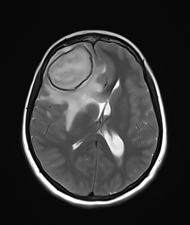
Thick-walled peripherally enhancing lesion measuring approx. 45 x 45 x 38 mm (AP x TD x CC) is seen in right frontal lobe. The lesion shows central diffusion restriction with low ADC. Associated mild dural enhancement is seen along right frontal convexity.
Hyperintensity with enhancement is seen involving frontal bone in midline, closely abutting anterior aspects of superior sagittal sinus.
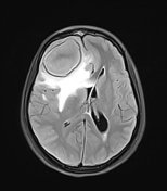
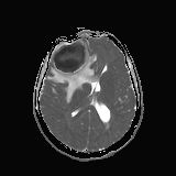
Gross perilesional oedema is seen extending to genu, anterior body of corpus callosum, right basal ganglia and external capsule with midline shift of approx. 14 mm towards left side and descending transtentorial herniation of right medial temporal lobe.
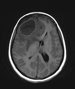
Compression of right lateral ventricle is seen with dilatation of posterior body, occipital horn of left lateral ventricle and periventricular seepage.
Bilateral anterior cerebral arteries are deviated towards left side.
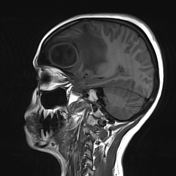
Mucosal thickening is seen in left frontal sinus with erosions / break in inner table, abutting adjacent left frontal lobe parenchyma. No obvious sizeable lesion is however seen in left frontal lobe.
Partially empty sella is seen with pituitary gland flattened against sellar floor – likely due to raised intracranial pressure.
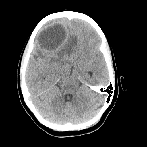
CT confirms the frontal bone osteomyelitis, left frontal sinusitis and break in its inner table.
Case Discussion
Right frontal lobe abscess with associated mild pachymeningitis and frontal bone osteomyelitis. The abscess shows diffusion restriction, T2 hypointense rim and 'dual rim sign' (well almost) on SWI. The source of infection is likely left frontal sinus.


 Unable to process the form. Check for errors and try again.