
Presentation
Memory disturbance.
Patient Data
Age: 70 years
Gender: Male
From the case:
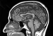
Cerebral amyloid angiopathy

Download
Info
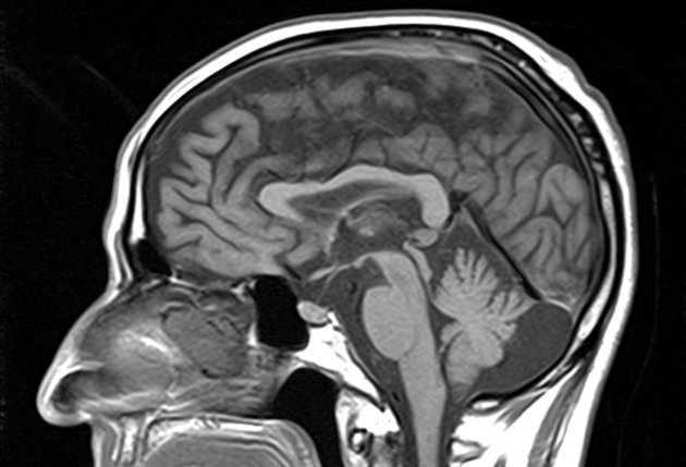
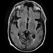
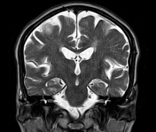
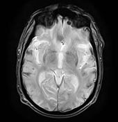
Multiple foci of susceptibility artifact are noted along the surface of the cerebellar/cerebral hemispheres and subarachnoid space of the cerebral convexity suggestive of micro-haemorrhages. Note sparing of the basal ganglia and pons.
White matter T2/FLAIR hyperintense lesions mainly involve the periventricular regions and centrum semiovale (chronic small vessel ischaemia).
Case Discussion
MRI features of multiple areas of susceptibility artifact in the cerebellum and cerebral hemispheres (micro haemorrhage) with convexity subarachnoid haemorrhage suggest probable cerebral amyloid angiopathy.
The main differential diagnosis is the hypertensive microangiopathy which is characterised by
- haemorrhages, including micro haemorrhages, typically located in basal ganglia, pons and cerebellum
- not associated with subarachnoid haemorrhage or superficial siderosis


 Unable to process the form. Check for errors and try again.