
Presentation
Headache of several months duration, nausea and vomiting. Heavy smoker in her youth.
Patient Data
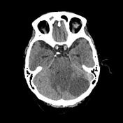


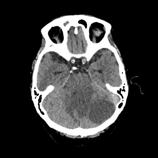
Cystic/necrotic nodule in the right occipital lobe with irregular thin enhancing walls and central hypoattenuation (30 HU).
Left cerebellar cystic/necrotic mass with well-defined walls and enhancing nodular component.
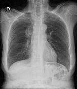
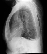

Right parahilar mass, with a "hidden hilum" sign.

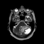
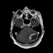
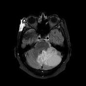
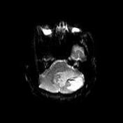
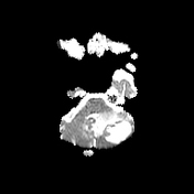
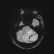
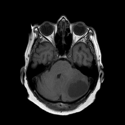
The multilobulated cystic lesion in the left cerebellar hemisphere shows high internal T2 signal that is partially suppressed on FLAIR and has thin nodular borders that enhance with contrast medium. On VEN BOLD sequence it shows peripheral areas with absent signal.
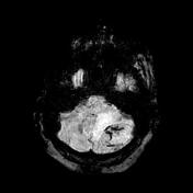
There is a small lesion with similar characteristics in the right occipital lobe. On DWI there is a small focus of diffusion restriction in the border of the posterior fossa lesion that enhances with contrast medium. The lesion is associated with surrounding vasogenic oedema and mass effect.
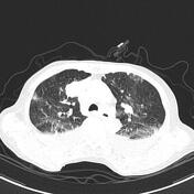
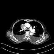

Heterogeneous lobulated nodule at the level of the right hilum with heterogeneous contrast enhancement occluding a branch of the right upper lobe.
Right hilar lymphadenopathy.
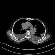
Small thrombus in the SVC.
Left pulmonary embolus with distal infarction.
Bilateral effusion with compressive atelectasis.
Case Discussion
The patient underwent craniotomy with removal of the left posterior fossa mass which was confirmed to be metastatic non-small cell lung cancer (NSCLC) on histology.
Multiple cystic/necrotic intracranial lesions with haemosiderin rim, proteinaceous content and enhancing solid components in an older adult are commonly due to metastatic disease; lung cancer is a common primary site.
Squamous cell cancer in the lung is often centrally located and necrotic. They commonly metastasise to brain.


 Unable to process the form. Check for errors and try again.