
Presentation
Patient presented with insidious progressive dementia.
Patient Data


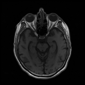
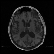
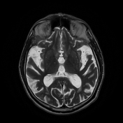
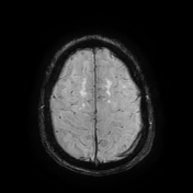
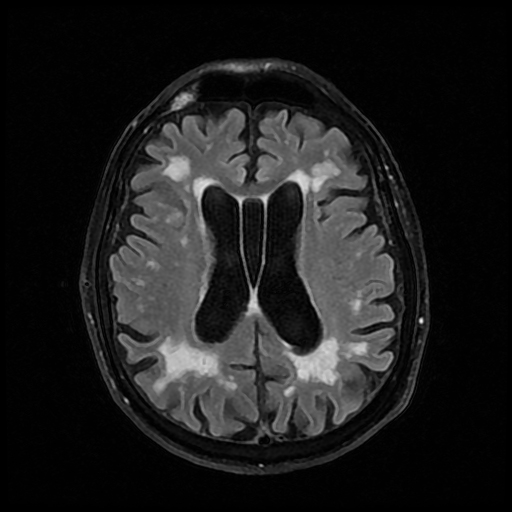
Extensive areas of leukoaraiosis on FLAIR and T2 sequences is noted in a subcortical and periventricular white matter distribution. The most prominent areas of involvement are adjacent to the anterior horn and occipital horn of the lateral ventricles. There is no evidence of hemorrhage or infarction. There is no evidence of hemosiderin deposition on susceptibility weight imaging to suggest chronic bleed or amyloid deposition.
Generalized atrophy of the cerebrum is noted causing a prominent sulcal pattern and ex vacuo enlargement of the ventricular system. There is no evidence of hydrocephalus. Incidentally noted is persistence of the septum pellucidum.
The cerebellum, brainstem, and cephalic portion of the spinal cord are well maintained.
The cranial nerves are unremarkable.
The basilar, internal carotid arteries and circle of willis are without evidence of aneurysm or occlusion.
Case Discussion
Cerebral small vessel disease is most often due to arteriosclerosis and are seen in a large portion of the population. It is considered the most common chronic and progressive vascular disease. While the findings are not always symptomatic they can lead to gait disturbances, depression, and cognitive impairment.
The differential diagnosis for cerebral microangiopathy is often cerebral amyloid angiopathy. The latter which more commonly affects the cortical portion of the cerebrum and is characterized by hemosiderin deposits.


 Unable to process the form. Check for errors and try again.