
Presentation
Short neck with restricted neck motions.
Patient Data
Age: 16 years
Gender: Female
From the case:
Chiari I with syrinx and skull base anomalies

Download
Info

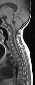
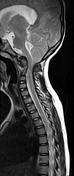
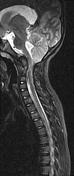
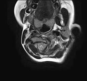
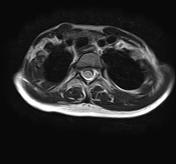
The MRI sequences demonstrate:
- caudal descent of the cerebellar tonsils through the foramen magnum 18 mm below the McRae line and 12 mm below the McGregor line.
- dilatation of the supratentorial ventricular system and 4th ventricle.
- skeletal anomalies with basilar invagination (the tip of the odontoid process projects 2 mm above the McRae line and 7 mm above the McGregor line) with compression of the bulbomedullary junction.
- intramedullary cystic cavity of the upper dorsal spinal cord in keeping with syrinx.
From the case:
Chiari I with syrinx and skull base anomalies
Download
Info

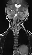
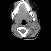
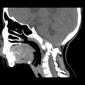
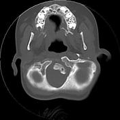
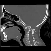
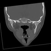
The CT scan demonstrates:
- dextroscoliosis.
- hypoplasia of the right occipital condyle.
- midline defect of the anterior arch of the atlas, with agenesis of the posterior arch.
- hypoplasia of the lateral masses of C1 (more prominent on the left)
- hypoplasia of the odontoid process and the body of C2.
- basilar invagination compressing the bulbomedullary junction.
- vertebral block from C2-C3 to C4-C5 level (Klippel-Feil malformation type 2)
Case Discussion
CT and MRI features of a Chiari I malformation with associated hydrocephalus, syrinx and skeletal anomalies.
Additional contributor: ZE. Boudiaf, MD CHU, Constantine, Algeria


 Unable to process the form. Check for errors and try again.