
Presentation
Abdominal discomfort.
Patient Data


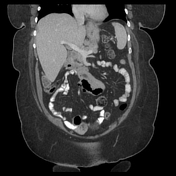
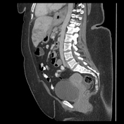
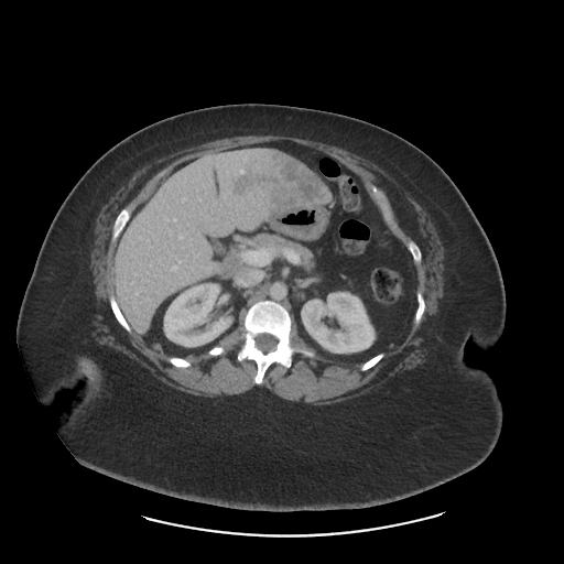
Large, heterogeneously enhancing mass in the left hepatic lobe, which has broad areas of hypoenhancement and peripheral areas of hypoenhancement. The extent of the mass can be best appreciated on coronal images, where it has an infiltrative component that extends laterally towards the fissure, with associated thrombosis of the anterior segment 2/3 branch of the portal vein. No other liver masses. Single enlarged gastrohepatic lymph node.
Case Discussion
Mass-forming intrahepatic cholangiocarcinoma, with associated thrombosis of the segment 2/3 portal vein. At first glance, particularly on the axial images, this could be mistaken for a hemangioma, as there is some element of peripheral discontinuous enhancement, however, note is the rind of enhancement does have a continuous component and the mass also appears quite large and infiltrative on the coronal images. Additionally, thrombosis of the portal vein is not a benign feature and concerning for either hepatocellular carcinoma, cholangiocarcinoma, or a bi-phenotypic type tumor. The other overlapping diagnosis to consider would be an abscess, however, noted that there is no surrounding edema in the liver parenchyma, and the degree of heterogeneous enhancement is not typical for an abscess. In this case, the next appropriate breast up would be an ultrasound-guided biopsy for pathologic confirmation, which was performed in this case and confirmed cholangiocarcinoma.


 Unable to process the form. Check for errors and try again.