
Presentation
Abdominal discomfort
Patient Data
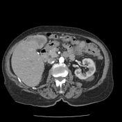

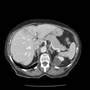

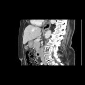
Large, heterogeneously enhancing mass within the anterior right hepatic lobe predominantly involving hepatic segments 4A and 4B. This is associated with some irregularity of the contour of the liver. There are small rounded lesions along the superior aspect marginating hepatic segment 8 and inferiorly near hepatic segment 5 which could reflect small satellite lesions. No lesions elsewhere. No intrahepatic biliary ductal dilation. The gallbladder is absent.
No adenopathy or other significant findings.
Case Discussion
Typical imaging findings of mass-forming intrahepatic cholangiocarcinoma, which was confirmed pathologically. At first glance, while there is some heterogeneous enhancement that could make you think about hemangioma, there is no peripheral nodular enhancement or other typical features, and the mass is quite large and irregular. Thus you now consider other liver lesions such as primary liver tumor, abscess or metastasis.
For abscess, there is usually a supporting clinical picture and more fluid attenuation of the lesion with surrounding edema. Metastases are usually multiple rather than a large dominant mass like this. Finally, for HCC, note that there is no avid arterial enhancement or characteristic washout, rather the mass retains heterogeneous enhancement on both phases. Thus, cholangiocarcinoma becomes the most likely diagnosis and can be confirmed with ultrasound-guided biopsy as the next step, as there is no biliary obstruction to guide management towards endoscopy.


 Unable to process the form. Check for errors and try again.