
Presentation
Right conductive hearing loss and otorrhoea.
Patient Data
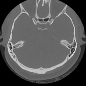

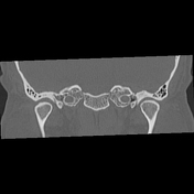
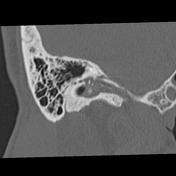
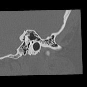

Filling of the right tympanic cavity by soft tissue material, which surrounds the ossicles and extends from the epitympanum to the hypotympanum. This material slightly expands Prussak's space and partially erodes the scutum and the ossicles. However, no erosions of the tegmen tympani are observed.
Also, there is mastoid air cells opacification on the right side (chronic otomastoiditis).
As additional findings, there is mucosal lining thickening and occupation at the ethmoid air cells and sphenoid sinus by soft tissue material (chronic sinusitis).
There are no alterations in the left ear.
Case Discussion
Occupation of the tympanic cavity with soft tissue material, and the presence of erosive changes in the bony structures of the middle ear, in an adult patient, are findings suggestive of acquired cholesteatoma (98% of cases). The presence of ipsilateral otomastoiditis indicates that it is a secondary acquired cholesteatoma, and the expansion of Prussak's space suggests that it originates at the level of the pars flaccida of the tympanic membrane (the most common type) 1.
Differential diagnoses to consider are chronic otitis media, cholesterol granuloma and facial nerve schwannoma 1.
High-resolution CT (HRCT) scan of the temporal bone is an accurate method for cholesteatoma and successful identification of ossicular status, location and extent of disease 2. However, HRCT temporal bone has some limitations like differentiating cholesteatoma from a chronic inflammatory process, especially in early disease states where there is an absence of osseous erosion. Also, the integrity of the facial canal cannot be accurately evaluated by HRCT. Because of the acquisition of the non-contrast CT, intracranial complications are often dismissed 1.


 Unable to process the form. Check for errors and try again.