
Presentation
Left side slowly progressive hemifacial deformity and pain with left eye proptosis for almost one year.
Patient Data
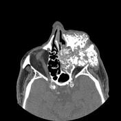


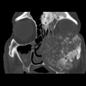
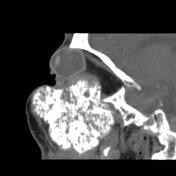
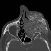

A large lytic-expansile cauliflower growth mass lesion with bone destruction and numerous polymorphic ossified and calcified foci axial and size with axial width up to 70 x 65 mm and height up to 60 mm in left maxillary bone extended within left orbital and nasal cavity is seen which is also bulged within the left cheek and buccal space and also retromaxillary and pterygopalatine fossa. The mass compressed the left eye onto the left orbital roof and also extended within the left zygoma bone body and maxilla alveolar process, and infratemporal fossa.
The right eye has no native lens and has exodeviation. Small calcified focus in the anterior temporal side uveo-scleral layer of both eyes in the insertion of lateral rectus muscle tendon is seen.
Case Discussion
The case illustrates the non-contrast MDCT features of pathology-proved low-grade chondrosarcoma of the maxilla. Head and neck chondrosarcoma are relatively rare but the maxillary bone is one of the major locations of the tumor and in most of the cases are low-grade and the main treatment for the tumor is wide local excision 1.


 Unable to process the form. Check for errors and try again.