Choroid plexus papilloma
Updates to Case Attributes
CT and MRI of the brain of a 4 year old demonstrates a mass in the trigone of the left lateral ventricle. The mass is relatively well circumscribed, slightly hyperdense compared to grey matter on CT, and following administration of contrast demonstrates vivid enhancement.
On MRI the mass is near-isodense to grey matter on T1 weighted sequences, heterogeneous and hyperdense on T2 and also demonstrates vivid contrast enhancement.
Location and appearances are consistent with a choroid plexus papilloma, subsequently confirmed on histology.
-<p>CT and MRI of the brain of a 4 year old demonstrates a mass in the trigone of the left lateral ventricle. The mass is relatively well circumscribed, slightly hyperdense compared to grey matter on CT, and following administration of contrast demonstrates vivid enhancement. </p><p>On MRI the mass is near-isodense to grey matter on T1 weighted sequences, heterogeneous and hyperdense on T2 and also demonstrates vivid contrast enhancement. </p><p>Location and appearances are consistent with a <a href="/articles/choroid-plexus-papilloma-1" title="Choroid plexus papilloma">choroid plexus papilloma</a>, subsequently confirmed on histology. </p>- +<p>CT and MRI of the brain of a 4 year old demonstrates a mass in the trigone of the left lateral ventricle. The mass is relatively well circumscribed, slightly hyperdense compared to grey matter on CT, and following administration of contrast demonstrates vivid enhancement. </p><p>On MRI the mass is near-isodense to grey matter on T1 weighted sequences, heterogeneous and hyperdense on T2 and also demonstrates vivid contrast enhancement. </p><p>Location and appearances are consistent with a <a href="/articles/choroid-plexus-papilloma-1">choroid plexus papilloma</a>, subsequently confirmed on histology. </p>
Updates to Study Attributes
The mass is relatively well circumscribed, slightly hyperdense compared to grey matter on CT, and following administration of contrast demonstrates vivid enhancement.
Image CT (C+ delayed) ( update )

Image CT (non-contrast) ( update )

Image 1 CT (non-contrast) ( update )
Image 2 CT (C+ delayed) ( update )
Updates to Study Attributes
On MRI the mass is near-isodense to grey matter on T1 weighted sequences, heterogeneous and hyperdense on T2 and also demonstrates vivid contrast enhancement.
Image MRI (T2) ( update )
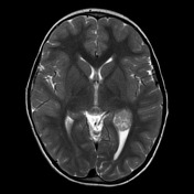
Image MRI (FLAIR) ( update )
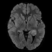
Image MRI (DWI) ( update )

Image MRI (T1) ( update )

Image MRI (T1 C+) ( update )
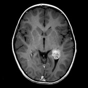
Image MRI (T1 C+) ( update )
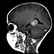
Image MRI (T1 C+) ( update )
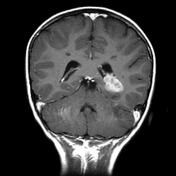
Image 1 MRI (T2) ( update )
Image 2 MRI (FLAIR) ( update )
Image 3 MRI (DWI) ( update )
Image 4 MRI (T1) ( update )
Image 5 MRI (T1 C+) ( update )
Image 6 MRI (T1 C+) ( update )
Image 7 MRI (T1 C+) ( update )