
Presentation
Headache, nausea and vomiting.
Patient Data
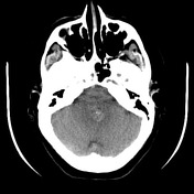

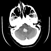

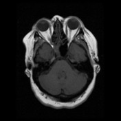
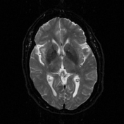
A vividly enhancing mass in the 4th ventricle is present, with speckled calcification. It is a solitary lesion, and no hydrocephalus is present.
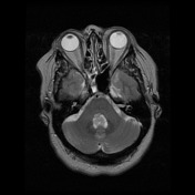
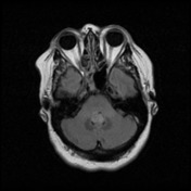

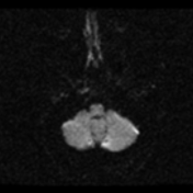
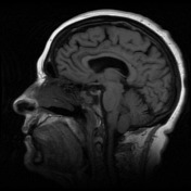
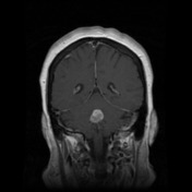
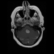

A 17 x 16 x 17 mm mass is seen within the floor of the fourth ventricle. No extension into the foramen of Luschka. It is isointense to parenchyma on T1, T2 hyperintense, and mildly hyperintense on FLAIR, with vivid diffuse contrast enhancement. No diffusion restriction. No foci of haemorrhage seen on EPI, although there are areas of linear hyperdensity on CT that may represent calcification.
No hydrocephalus seen.
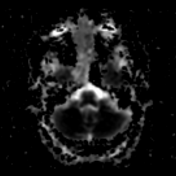
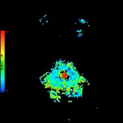
Perfusion study demonstrates increased cerebral blood flow within the mass. MR spectroscopy (not shown) over the mass demonstrates slight reduction in NAA and reversal of the choline to creatinine ratio, although unfortunately a sloping baseline reduces specificity of findings.
T2/FLAIR white matter and periventricular hyperintensity in keeping with chronic small vessel ischaemic change. Fluid seen within the ethmoid and sphenoid sinuses.
Conclusion:
Tumour within the floor of the fourth ventricle. Given the CT and MRI appearances the differential includes ependymoma, more likely (given the vivid enhancement) a choroid plexus papilloma. Less likely possibilities include a metastasis, or even less likely intraventricular meningioma.
Case Discussion
The patient went on to have a resection.
Histology
MICROSCOPIC DESCRIPTION: Paraffin sections confirm the frozen section diagnosis of choroid plexus papilloma. The consist of fragments of delicate papillary structures with fibrovascular cores covered by a single layer of cytologically unremarkable choroid plexus epithelium. No mitotic figures, solid papillae or areas of necrosis are identified.
DIAGNOSIS:


 Unable to process the form. Check for errors and try again.