
Presentation
5 weeks of progressively worsening muscle weakness and decreasing muscle tone.
Patient Data
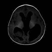


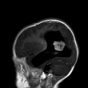
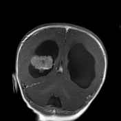
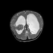
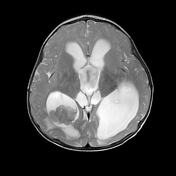
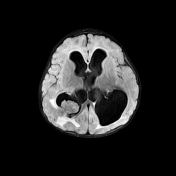
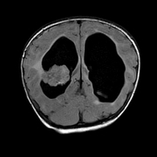
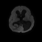
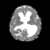

There is a well-circumscribed, homogeneously enhancing, lobulated mass within the trigone of the right lateral ventricle that displays an intermediate T2 signal. The lateral margin of the mass is contiguous with the subependymal surface of the right lateral ventricle. Furthermore, there is dilatation of the supratentorial and infratentorial ventricular system with secondary cephalad bowing of the corpus callosum.
Case Discussion
This is a case of a choroid plexus carcinoma of the right lateral ventricle. The patient underwent excision of the right intraventricular tumour.
Histology
Histopathologic analysis revealed that the carcinomatous foci exhibited solid growth, increased mitotic activity (>4 mitoses per 1 field under 40x lens in multiple foci), and necrosis.
Stains of the tumour were positive for AE1/AE3 and Cam 5.2, with focal staining for GFAP, Syn, and vimentin. In addition, the tumour showed elevated Ki-67 (30% - 40% in many foci) and p53 (>50% in many foci), with limited, focal staining for S-100 and SOX-10.


 Unable to process the form. Check for errors and try again.