
Presentation
Migraine, associated with neck pain and tinnitus for 9 months.
Patient Data


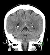


No CT evidence of recent major territorial infarction or intracranial hemorrhage. Prominent choroid plexuses of both lateral ventricles, with displacement of choroidal calcifications around their periphery. Empty sella.


Choroid plexus lesions in the trigone bilaterally. These lesions are close to CSF on T1 & T2-weighted images but don’t attenuate completely on FLAIR, high signal on DWI with intermediate ADC signal and don’t enhance. Enlarged sella/empty sella.
Case Discussion
CT & MRI findings are suggestive of bilateral choroid plexus xanthogranulomas. Its an asymptomatic incidental finding. The neurological evaluation for the empty sella/enlarged sella was also unremarkable.


 Unable to process the form. Check for errors and try again.