
Presentation
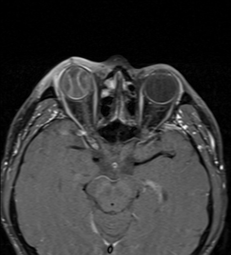
Corneal, scleral, and upper eyelid lacerations have been sutured, with a suspected intraocular foreign body following injury caused by a grinding wheel fragment.
Patient Data
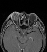

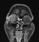
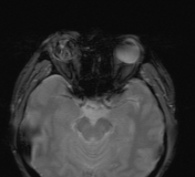
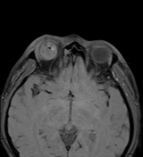

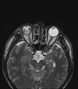
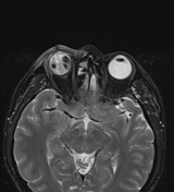
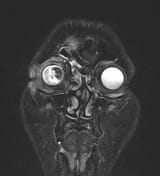
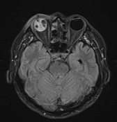
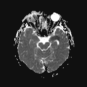
Bilateral choroidal detachment in the right eye with convex intraorbital leaflets extending anteriorly beyond the ora serrata reference point, along with choroidal haemorrhage.
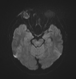
A low signal nodule on multiple sequences in the vitreous cavity suggests a possible foreign body.
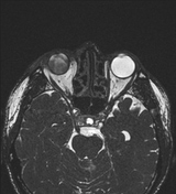
The shape and signal of the lens are not visible.
The right eyeball is distorted and slightly smaller compared to the left. Heterogeneous fluid in the vitreous cavity, mildly low signal on GRE, likely represent haemorrhage.
Soft tissue oedema in the upper eyelid with a skin discontinuity, which has been sutured.
Case Discussion
The imaging findings are consistent with choroidal detachment with choroidal haemorrhage, an intraocular foreign body, lens trauma, and oedema with a laceration of the upper eyelid skin.


 Unable to process the form. Check for errors and try again.