
Presentation
Day 2 post-op penetrating keratoplasty (corneal transplant) OD. Right hemiparesis, frequent falls towards his left side. Rule out stroke.
Patient Data
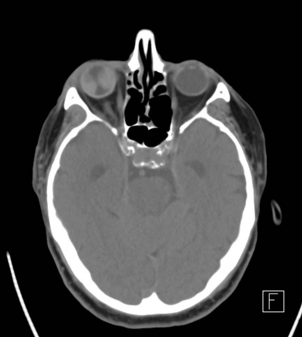
1- in reference to the clinical question, there are no notable acute ischaemic lesions
2- hyperdense lentiform lesion along the lateral and medial margins of the posterior segment of the right globe. Sparing of the most posterior portion of the globe
3- aphakia OD (as confirmed by the operative report)
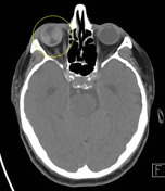
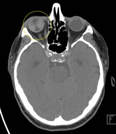
Hyperdense lentiform lesions, compatible with haemorrhage.
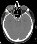
Notice how the detachment is not limited anteriorly by the ora serrata (inferred reference point). Also, posteriorly, the detachment diverges at the optic disc.
Case Discussion
Here's how to differentiate choroidal versus retinal detachment:
The main difference is that fluid and blood that accumulates within the sub-choroidal space diverge at the optic disc.
Whereas fluid and blood that accumulates between the retina and choroid layers (thus resulting in retinal detachment) will manifest as a V-shaped fluid lesion in the posterior segment of the globe, with the apex at the optic disc. Posteriorly, the detachment converges on the optic disc.
There are different aetiologies of choroidal haemorrhage. Here, it occurred peri-operatively.


 Unable to process the form. Check for errors and try again.