
Presentation
Progressive dyspnea developing over three weeks subsequent to viral illness. Associated with productive cough, fatigue, malaise, and night sweats. Inflammatory markers elevated. Peripheral eosinophils > 7.0 x10*9/L
Patient Data

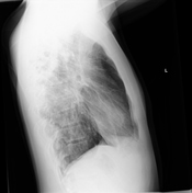
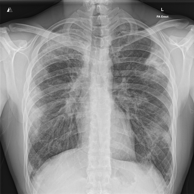
Multifocal patchy consolidation.
Peripheral distribution.
Cardiac and mediastinal contours normal.
No pleural effusion.

Bilateral peripheral multifocal patchy airspace consolidation and ground-glass opacification with upper zone predominance.
Multiple air bronchograms.
Moderate enlargement of subcarinal, pre-tracheal, and hilar lymph nodes.
No solid mass lesions. No pleural effusion.

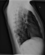
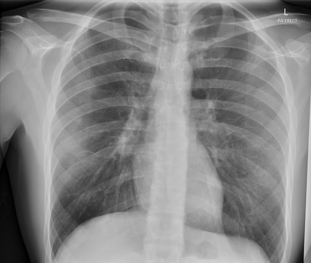
Substantial improvement compared with previous study.
Case Discussion
The patient presented due to exertional dyspnea developing over three weeks subsequent to a viral illness.
Other associated symptoms included productive cough, fatigue, myalgias, and night sweats.
The patient had a history of mild asthma and ankylosing spondylitis. There were no relevant environmental or drug exposures.
A full blood count showed a significant eosinophilia. The serum IgE was elevated to 700-800 IU/ml.
A subsequent CT chest showed multifocal peripheral airspace consolidation and ground-glass opacification, air bronchograms, and moderate enlargement of subcarinal, pre-tracheal, and hilar lymph nodes.
The patient was treated with oral corticosteroids and returned to normal function.


 Unable to process the form. Check for errors and try again.