
Presentation
Chronic left arm pain. History of surgery 1 month back for debridement of chronic osteomyelitis.
Patient Data
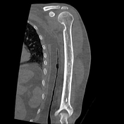


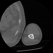
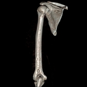
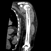
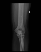

Diffuse thickening and remodelling of the humeral shaft. Small healing cloaca at the distal humeral shaft.
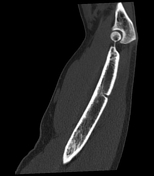
Another larger more distal surgical track, with a sclerotic bone fragment seen within (bony sequestrum).
Case Discussion
The patient presented for follow-up after surgical debridement 1 month back for chronic osteomyelitis.
He had presented 3 months earlier at another hospital with chronic pain of the left arm for 2 months, not responding to analgesics; No history of trauma, significant medical illness or comorbidities.
X-ray and CT (old studies in this case) and lab tests were done. The patient received IV antibiotics and was submitted for surgical debridement where histopathological verification of osteomyelitis was achieved.
The newly developed bony sequestrum may act as a nidus for infection and hence usually requires excision if a cure is to be achieved.


 Unable to process the form. Check for errors and try again.