
Presentation
Palpable abdominal mass
Patient Data
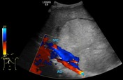
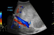


Large heterogenous abdominal mass exerting mass effect on both intraperitoneal and retroperitoneal structures. Although origin cannot be confidently determined, the lesion appears to be centred on right renal artery (which shows patent flow on spectral Doppler), suggesting a primary renal tumour.
Recommend further characterisation by CT.


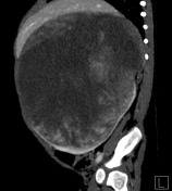
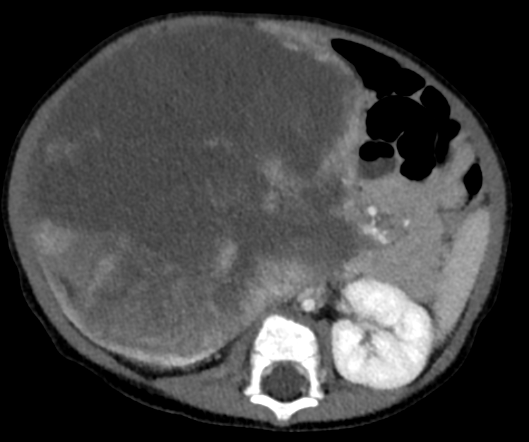
- large central mass massively expands right kidney, with a small amount of normal renal parenchyma is apparent on the periphery (claw sign)
- large central hypodense areas suggest necrosis
- no evidence of extra-renal invasion (limited assessment)
- pronounced mass effect on all adjacent structures
- compressed inferior vena cava, bowel
- mild upward displacement of liver
- small umbilical hernia
- no evidence of metastatic disease
(Presumably) on the basis of epidemiology, presumptive diagnosis of Wilms tumour was made. Patient was managed by initiation of neoadjuvant chemotherapy...

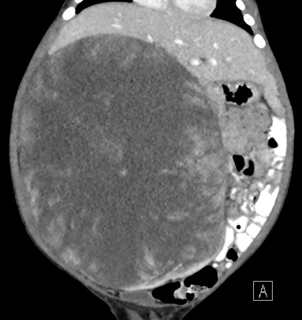
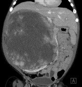
- interval enlargement of the mass, best appreciated by...
- further extension across midline (axial images)
- increased mass effect on liver (coronal image)
- decreased, only thin crescent, of normal right kidney parenchyma remaining
- there remains no evidence of extra-renal invasion or metastasis
Surgical pathology
Impression
Kidney and adrenal, right, radical nephrectomy
- Clear cell sarcoma of kidney, 19.8 cm
- tumours invades through capsule into perinephric fat and renal sinus lymphvascular spaces (stage ii)
- renal vein, ureteric and parenchymal resection margins free of tumour
Lymph nodes, paraaortic, excision
- No evidence of malignancy in two lymph nodes
--------------------------------------------------------------------------------------------------------
Microscopic description
- sections of renal mass show a monomorphic population of rounded cells arranged in a diffuse-to-corded pattern consisting of ovoid-to-round nuclei, vesicular chromatin, inconspicuous nucleoli. Cytoplasm is moderate, and mostly clear-to-eosinophilic. A delicate arborizing-to-parallel vascular network separates groups of tumour cells
- tumour-renal parenchymal interface is well-demarcated, focally infiltrating without pseudocapsule
- limited panel of immunostains show WT-1 to be negative in the tumour cells. INI-1 is retained. Cytokeratin fails to identify tubular cell rests and is negative
- overall, histomorphology is consistent with a clear cell sarcoma of the kidney, supported by its immunophenotype. By invasion through the capsule into the perinephric fat, as well as by invasion into the renal sinus lymphovascular spaces, this is stage II. Tumour is not seen present at ink, on multiple sections, therefore, presumed to be completely resected
Gross description
- first specimen labelled "right kidney and adrenal" consists of a previously inked and bivalved kidney and adrenal gland
- adrenal gland measures 6.5 x 1.5 x 0.1 cm. The gland is compressed on the surface of the kidney, but tumour does not grossly appear to invade it
- [right kidney] external surface is intact and glistening and not grossly disrupted. Cut surface appears heterogenous with tan-pink area mixed with regions of yellow-white friable parenchyma with regions of necrosis. Overall architecture is lobulated with regions of white-tan tissue having a glistening, mucoid appearance
- a green cassette labelled "ureter margin" and holds a tan-brown fragment of tissue measuring 1.6 x 0.6 x 0.7 cm and is compressed at one end
Case Discussion
This case highlights the ambiguity of solid paediatric renal masses, as well as some important points regarding the management of Wilms tumour.
By imaging appearance, solid paediatric renal tumours often appear similar and cannot be distinguished with high specificity. By epidemiology, a diagnosis of Wilms tumour is reasonable, as they account for ~90% of paediatric renal masses. However, uncommon lesions should be considered if...
- aggressive appearance (large amounts of necrosis)
- atypical age range (Wilms tumour peak incidence is 3 years old, never in newborns)
- other suggestive characteristic
- rhabdoid tumour: subcapsular fluid collection, substantial necrosis +/- linear calcifications
- clear cell sarcoma of kidney: bone metastases
Regarding management of Wilms tumour, there are two major entities which advocate different approaches:
- National Wilms Tumour Study / Children's Oncology Group (COG) - North America
- early surgical resection and staging, followed by chemotherapy
- International Society of Paediatric Oncology (SIOP) - Europe, South America
- neoadjuvant chemotherapy, followed by surgical resection and diagnostic confirmation
Although treatment outcomes are felt to be similar, one risk of the SIOP-type approach is the misdiagnosis of benign or malignant lesions based on imaging, since histopathologic confirmation may be delayed until the time of resection.
Due to rarity, there is no standard treatment for either rhabdoid tumour or clear cell sarcoma of the kidney 2.


 Unable to process the form. Check for errors and try again.