
Presentation
This patient was admitted to the emergency room as a motor vehicle accident victim, evolving with vomiting, drowsiness, and disorientation.
Patient Data


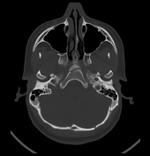
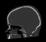
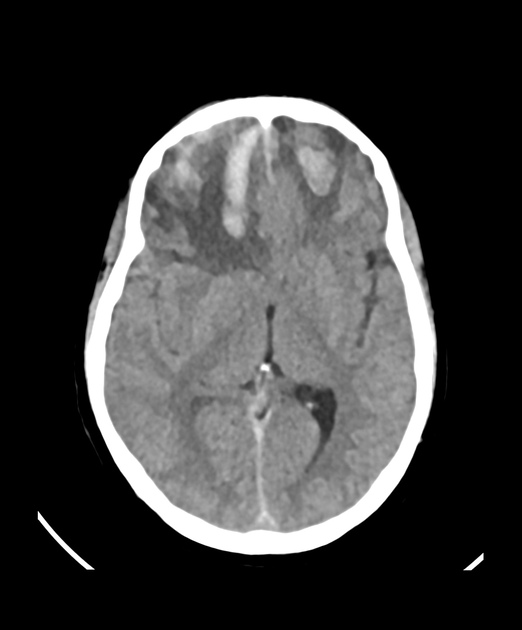
CT scans demonstrate a centrally transverse fracture line extending through the basiocciput (clivus) associated with occipital skull vault fractures. CT also reveals right frontal subdural haematoma, left parietal subarachnoid haemorrhage, haemorrhagic contusions in frontal lobe bilaterally, brain oedema, and sphenoid haemosinus.
Impression: Traumatic brain injury with skull vault fractures and a transverse clival fracture, associated with intracranial haemorrhage.
Case Discussion
Clival fractures are uncommon skull base fractures 1-5. CT pattern has predictive value about prognostic; therefore, radiologists working in the emergency department should be able to identify the types of fractures of the clivus and alert clinicians to their complications 1-5. This case illustrates a high-energy head trauma with a transverse clival fracture, a fracture of the occipital bone, and an intracranial haemorrhage.


 Unable to process the form. Check for errors and try again.