
Presentation
Abdominal pain.
Patient Data
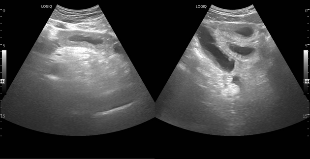
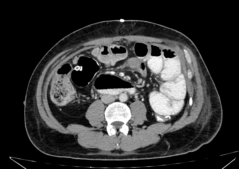
Dilated some bowel loops at the lower part of the abdomen with diffuse mural thickening.


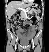
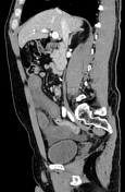
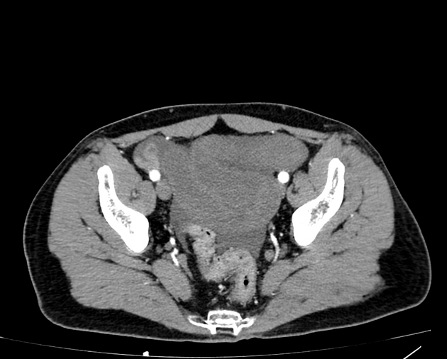
A cluster of dilated ileal bowel loops is seen in the pelvis with two adjacent transition points (in axial plane: proximal, distal), and (in sagittal plane: proximal, distal). There is diffuse thickening and hypoenhancement of the obstructed bowel loop reflecting ischemic changes. Mild free ascites is noted. Normal opacification of mesenteric arteries and veins. No portal venous gas and no pneumoperitoneum.


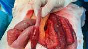

The patient was operated on after failed conservative management. Release of omental adhesive band and hot fomentation for the ischemic bowel were done. No bowel resection needed.
The last image is at one of the transition points and shows the difference in color between normal bowel and ischemic bowel.
Photoes courtesy Dr. Mahmoud Abu-elmakarem, consultant of surgery.
Follow up one day after surgery shows normal caliber of bowel loops and mild mural thickening of the previously obstructed bowel loops.
Case Discussion
On CT, the presence of two adjacent transition points is consistent with closed loop bowel obstruction, known as double beak sign. It needs urgent surgical intervention as it is usually associated with bowel ischemia.


 Unable to process the form. Check for errors and try again.