
Presentation
History of large B-cell intraocular lymphoma. Increasingly severe headaches.
Patient Data


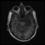
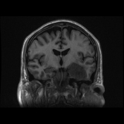
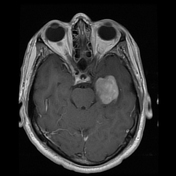
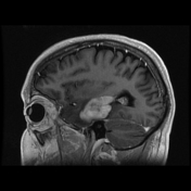
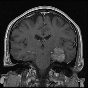
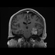
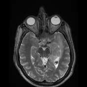
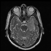
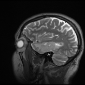
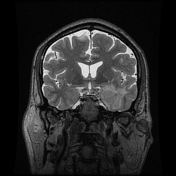
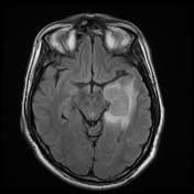
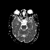
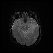
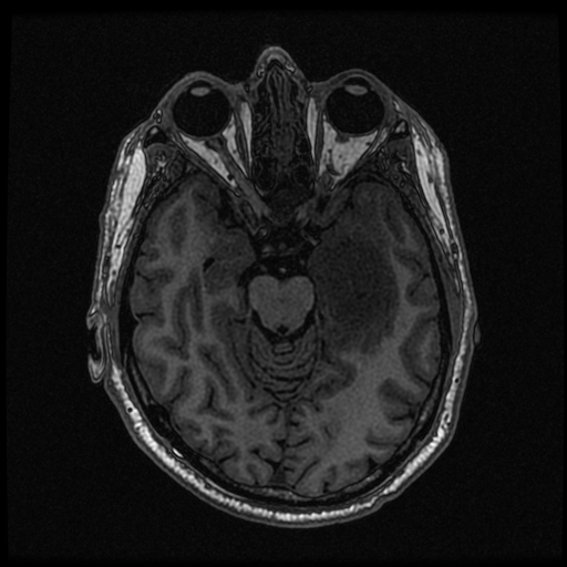
There is expansile intermediate T2 signal mass within the medial left temporal lobe, which
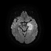
displays diffuse homogeneous enhancement. Furthermore, there is reduced diffusivity throughout the mass.
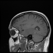
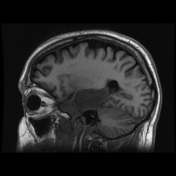
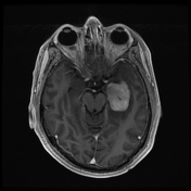
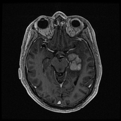
There is a homogeneously enhancing mass within the subcortical white matter of the left superior parietal lobe. Additionally, there are 3 nodular foci of enhancement within the medial right cerebellum.
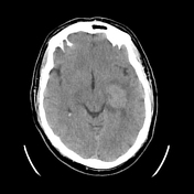
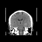
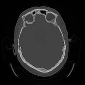
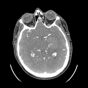
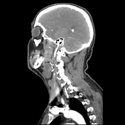
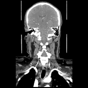
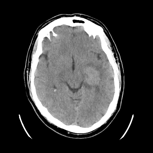
Redemonstration of an expansile lesion along the medial left temporal lobe. Additional lesions seen on MR are not visualised on this examination.
There is asymmetric prominence and increase in calibre of the left MCA vasculature, most noticeable within the M2 and M3 segments of the vasculature.
Case Discussion
This is a CNS lymphoma. The patient has a history of diffuse large B cell lymphoma. He was treated with several courses of rituximab, methotrexate, procarbazine, vincristine (R-MPV) and intraventricular rituximab.
Despite treatment, the patient had progressive right sided hemiataxia, dysarthria, and headaches. An MRI showed progression of right cerebellar lesion and concern for hydrocephalus due to ventricular involvement. He continued to have worsening mental status. He eventually expired due to complications related to therapy.
Co-author:
Jun Yang


 Unable to process the form. Check for errors and try again.