
Presentation
This patient presented with congenital deafness.
Patient Data
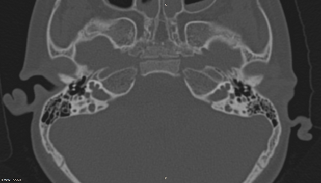
The cochlea is almost normal in size, but there is an incomplete partition. The modiolus is absent, and the interscalar septa are partially preserved, giving the cochlea a corkscrew appearance. There is no lamina cribrosa separating the cochlear base from a bulbous-appearing internal auditory canal fundus. The basal turn directly communicates with the internal auditory canal. The vestibular aqueduct is dilated. There is also a sac-like dilation projecting upward from the right utricle. Additionally, the otic capsule is thin.
Case Discussion
Cochlear incomplete partition type III (IP-III) is a rare, X-linked genetic disorder affecting the inner ear. It is characterised by a distinctive corkscrew-like appearance of the cochlea on imaging studies. In our patient, there is also a sac-like dilation projecting upward from the utricle. This anomaly results from the absence of the modiolus, the bony core that supports the cochlear structures.
Consequently, the cochlea lacks proper partitioning between its fluid-filled compartments, leading to mixed conductive and sensorineural hearing loss. While cochlear implantation is a potential treatment option, the anatomical abnormalities associated with IP-III can present significant surgical challenges and variable outcomes.


 Unable to process the form. Check for errors and try again.