
Colloid cyst of the third ventricle with obstructive hydrocephalus
Presentation
Bi-frontotemporal progressive headache for a few months, recurrent attacks of tonic colonic fits, associated with loss of consciousness, followed by prolonged sleep. Deterioration of memory over the past week.
Patient Data
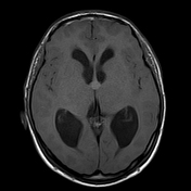

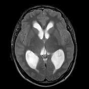
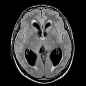
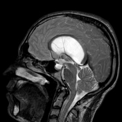
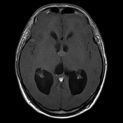
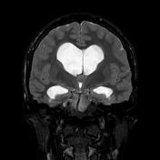
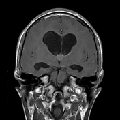
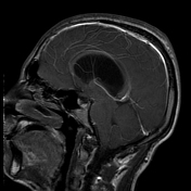

There is a midline well-defined cystic-like rounded lesion seen centred on the foramen of monro having iso to hyperintense signal on T1, low signal in T2, measures about 8 x 10 x 8 mm in diameter. There is associated moderate to marked symmetrical dilation of both lateral ventricles with periventricular bright T2 and FLAR band-like signal, likely related to CSF permeation.
Normal size of third and fourth ventricles.
Case Discussion
Classic appearance of colloid cyst of the third ventricle with acute obstructive hydrocephalus. In most cases, this lesion is incidentally discovered and usually asymptomatic. However, it can cause acute obstructive hydrocephalus which can be life-threatening. In this instance, craniotomy with excision via transcallosal or transcortical route, stereotactic aspiration, endoscopic removal, or external ventricular drain can be done 1.


 Unable to process the form. Check for errors and try again.