
Presentation
Acute left abdominal pain with tenderness. Known for numerous benign hepatic lesions.
Patient Data


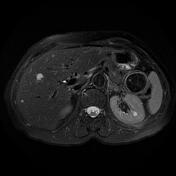
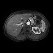
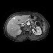
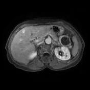
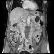
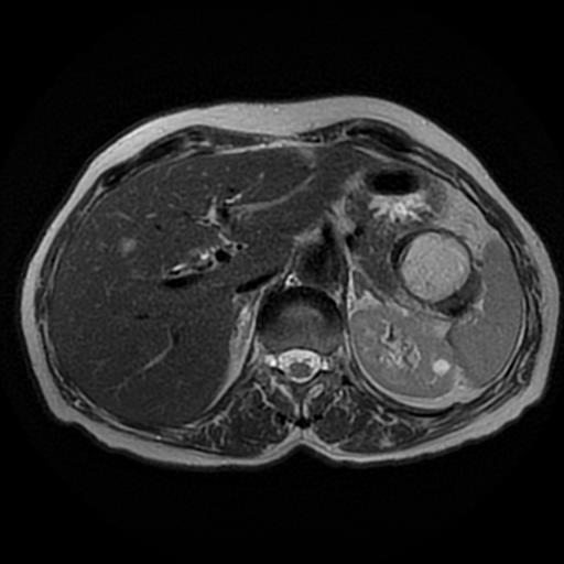
Numerous well-defined hepatic lesions of various sizes disseminated in both lobes, the largest lesion is located in segment 3 (22 mm). They display a low signal on T1, and a high signal on T2/T2 fat sat with no restricted diffusion. On postcontrast sequences, some of them show a quick, intense and homogeneous enhancement on the arterial phase (flash-filling haemangiomas), and others show a discontinuous nodular peripheral enhancement on the arterial phase, more centripetal fill-in on the portal venous phase with complete fill-in on delayed sequences.
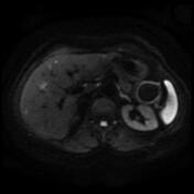
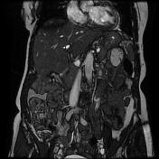
Evidence of colocolic Intussusception at the splenic flexure with a well-defined mass at the lead point of a high signal on T1, and T2, attenuated on fat sat sequences with no enhancement on postcontrast sequences, highly suggestive of a lipoma.
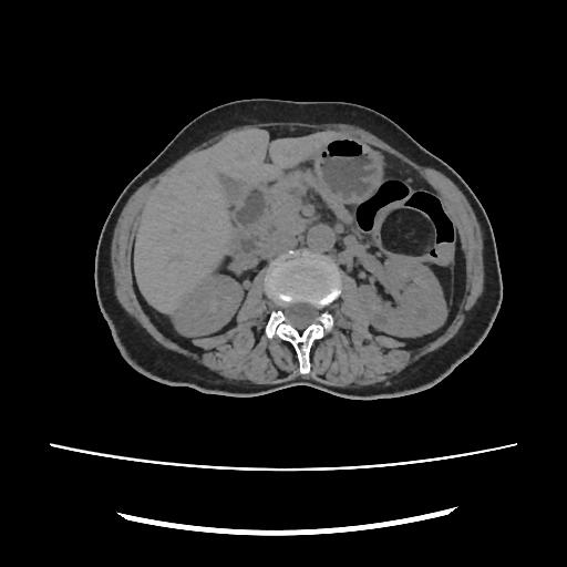
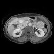
CT scan performed after the MRI exam showed the colocolic intussusception and fatty mass at the lead point.
Case Discussion
MRI and CT features of a colocolic intussusception secondary to a lipoma.
Gastrointestinal tract lipomas are not uncommon and can be found anywhere along the entire gastrointestinal tract. The colon is the most common location and is usually on the right side The vast majority are of submucosal location with only a small number of subserosal and can be sessile or pedunculated.
Usually, they are asymptomatic and found incidentally. They can be pedunculated and occasionally present as the leading point of an intussusception (as in this case). When large they may develop mucosal ulceration and present with iron deficiency anaemia or positive faecal occult blood testing.


 Unable to process the form. Check for errors and try again.