
Presentation
History of 11 weeks of amenorrhoea. Mild PV bleeding for 3 weeks, associated with nausea and vomiting. No fever.
Patient Data

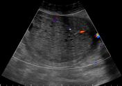
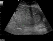
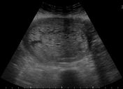

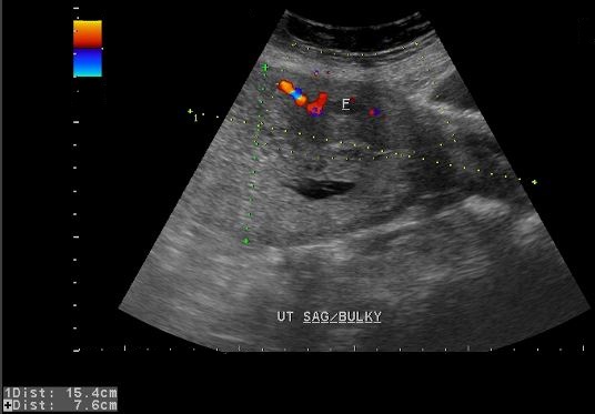
Bulky anteverted uterus with a large isoechoic intrauterine mass containing multiple tiny cystic areas (resembling snow storm or bunch of grapes). No significant internal vascularity is seen in it on colour Doppler ultrasound examination. No fetal parts are noted.
Impression: Sonographic features are suggestive of gestational trophoblastic disease (complete hydatidiform mole).

Histopathology report of the uterine contents removed during suction evacuation.
Case Discussion
Laboratory investigations demonstrated abnormally elevated serum beta HCG = 1037841 IU/L (normal range 0.0-5.0IU/L). The patient underwent dilation and suction evacuation. Histopathological analysis of the submitted specimen showed complete hydatidiform mole.


 Unable to process the form. Check for errors and try again.