
Presentation
Foot pain several weeks after an ankle fracture.
Patient Data


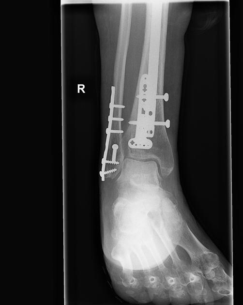
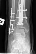
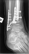
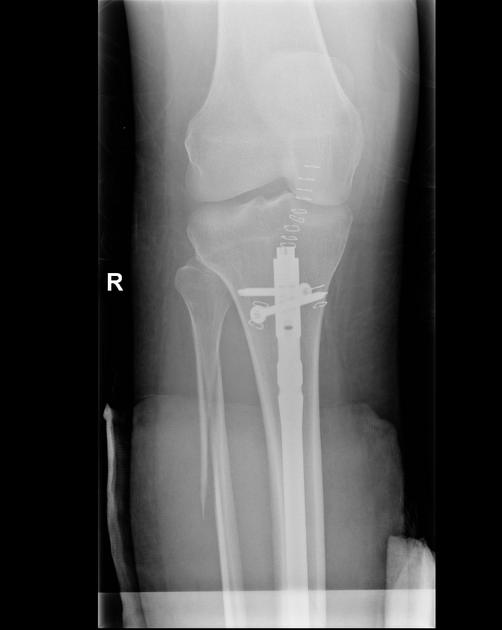
Right lower leg and ankle
Comparison is made with the previous examination several weeks earlier. Tibial intramedullary nail in unchanged position. There is delayed union of the oblique fracture through the distal tibial diaphysis. Screw and plate fixation of distal fibular fracture unchanged. There is non-union of the proximal right fibular spiral fracture. The ankle joint alignment is normal. No new fracture identified.
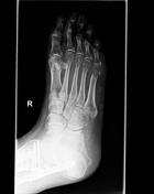
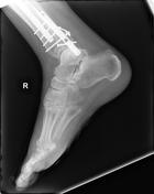
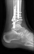
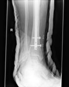
Right foot
There is severe patchy osteopenia predominantly in periarticular distribution. No new fracture identified. The overall appearances are most in keeping with reflex sympathetic dystrophy (Sudeck atrophy).
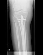

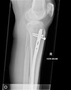
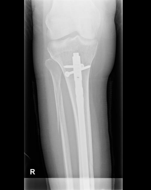
Tibial intramedullary nail transfixes a comminuted tibial shaft fracture.
New posterior distal tibial plate and lateral malleolar plate and screws.
Alignment at the mortise is within normal limits.
Minimally displaced proximal fibular fracture is unchanged in position.

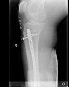
There has been no change in alignment of the intramedullary nail fixating a comminuted tibial diaphyseal fracture. Oblique fracture of the proximal fibular shaft is similar to previously.
Comminuted distal fibular fracture involving the lateral malleolus, displaced posterior tibial plafond fracture and minimally displaced medial malleolus fracture are similar to previously.
Case Discussion
Although there was no foot fracture, pain developed in the mid- and forefoot. Radiographs demonstrate severe regional osteopenia, typically in the periarticular regions, with appearances mimicking focal lytic bone lesions.


 Unable to process the form. Check for errors and try again.