
Presentation
Arrived to the ED by ambulance - obtunded, no reaction to verbal or tactile stimulation, tonic-clonic seizure. Hypertonic - no reliable way of checking for nuchal rigidity. Suppuration from right ear; known recurrent otitis media in same ear.
Patient Data
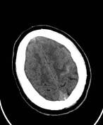

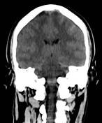


Left frontal subarachnoid hemorrhage (SAH).
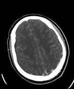
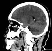
Diffuse cerebral edema, manifesting as sulcal and cisternal effacement.
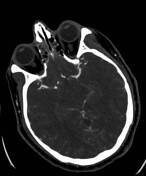
Perimesencephalic, suprasellar, right parafalcine, and bilateral Sylvian cistern pseudosubarachnoid hemorrhage, representing accentuated cerebral arteries due to said cerebral edema.
Thin parietal subdural collection on each side, right larger than left - most probably bilateral subdural empyema (SDE).
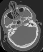
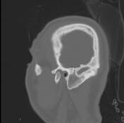
Air-fluid level in left maxillary sinus. Bilateral mucosal thickening in maxillary sinuses and most ethmoidal air cells. Mastoid air cells undeveloped on right and underdeveloped on left.
Opacified right middle ear and antrum. Adjacent minimal right temporal pneumocephalus, apparently due to dehiscence of the tegmen tympani, plus one right frontal parafalcine bubble.
Slightly prominent, enhancing adenoids, occluding the Eustachian tubes and fossae of Rosenmüller.
Non-occlusive thrombosis in posterior part of superior sagittal sinus (SSS).
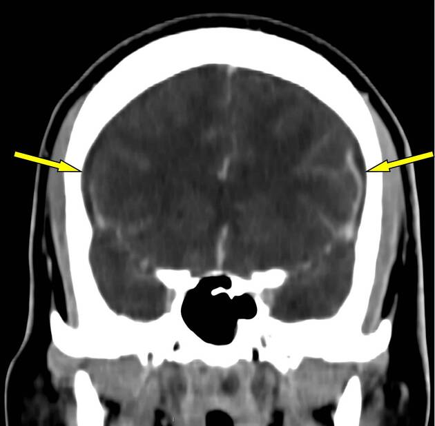
The arrows point to the subdural collections.
Case Discussion
A brain MRI (not shown) was promptly performed, verifying diffuse cerebral edema, bilateral subdural empyema, superior sagittal sinus (SSS) thrombosis, and left frontal subarachnoid hemorrhage (SAH).
The patient had been suffering from recurrent right otitis media, only this time it resulted in meningitis, plus the aforementioned complications. The dural venous sinus thrombosis was probably responsible for the SAH.
Blood cultures grew Streptococcus pyogenes.
Despite rigorous treatment, the patient passed away.


 Unable to process the form. Check for errors and try again.