Presentation
Recurrent loss of hearing and ear drainage in the left ear. History of resection of congenital cholesteatoma >10 years ago.
Patient Data
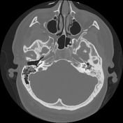

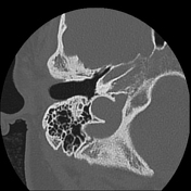

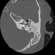

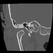

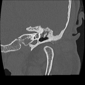

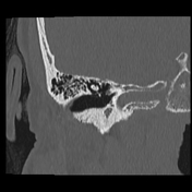

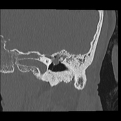

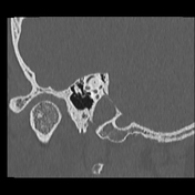

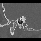

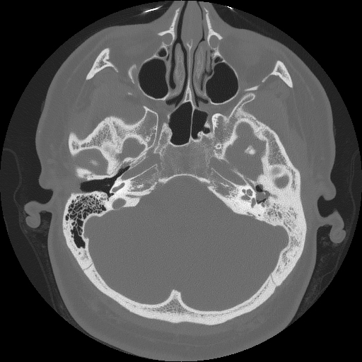
There is apparent focal defect involving the superior aspect of the tympanic membrane on the left. There is a left middle ear cavity soft tissue density involving the epitympanum (including Prussak's space) and mesotympanum. There is erosion of the left malleolar head, long process of the incus, and scutum. There is also erosion/demineralisation of the anterior aspect of the tegmen tympani on the left.
Case Discussion
This is a case of a recurrent congenital cholesteatoma. The patient originally had the mass resected 10-15 years ago as a child. Histopathology at that time revealed a tan/white mass with keratinaceous debris, consistent with a cholesteatoma.
The patient then began to experience recurrent hearing loss and ear drainage. The above CT of the temporal bones demonstrated findings highly suggestive of recurrence of her congenital cholesteatoma. She underwent resection of the mass which again showed a tan/white mass containing keratinaceous debris.
Co-author:
Mason Soeder


 Unable to process the form. Check for errors and try again.