
Presentation
Pain of the right hip with palpable mass at the superomedial aspect of the thigh.
Patient Data
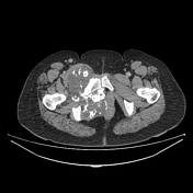

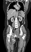
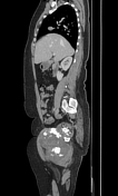

There is a large lobulated soft tissue mass centred on the right superior pubic ramus with mild and heterogeneous enhancement, containing multiple central rings and arcs calcification (cartilaginous matrix). It shows three components, two endopelvic components, one filling the right pararectal space and ischiorectal fossa with mass effect on the anorectal region which is displaced to the left and the other one anterior to the urinary bladder compressing and displacing the latter posteriorly against the uterus, the third component is of extrapelvic location, extending through the obturator foramen to the superomedial soft tissue of the right thigh. Small right inguinal lymphadenopathy is noted.
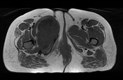
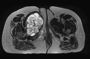
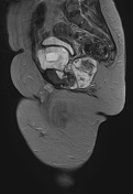
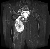
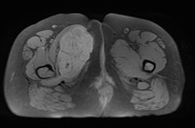
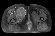
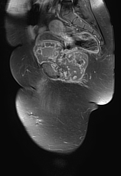
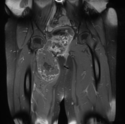
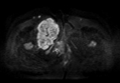
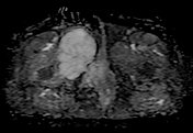

The previously described soft tissue mass appears lobulated multiloculated of low signal intensity on T1WI, heterogeneous high signal intensity on T2WI and STIR with heterogeneous enhancement on postcontrast sequences. The cartilaginous matrix demonstrates a low signal on T1WI with areas of high signal, and low signal T2WI. The obturator externus, pectineus, and adductor longus muscles are displaced anteriorly, and adductor brevis muscle posteriorly with most likely involvement of the adductor magnus muscle. The gracilis muscle is atrophied.
Case Discussion
CT and MRI features characteristic of chondrosarcoma, histologically-proven as conventional chondrosarcoma grade I according to O'Neal and Ackermann classification
Conventional chondrosarcoma, also known as central chondrosarcoma, is considered as the most common subtype of chondrosarcoma. It may be of low, intermediate or high grade. The low-grade conventional chondrosarcomas (as in this case) can be difficult to differentiate from an enchondroma.


 Unable to process the form. Check for errors and try again.