
Presentation
This patient presented to the ER with desaturation and tachypnoeic. He reports that he started with episodic fever, dyspnoea, cough, and odynophagia eight days ago.
Patient Data
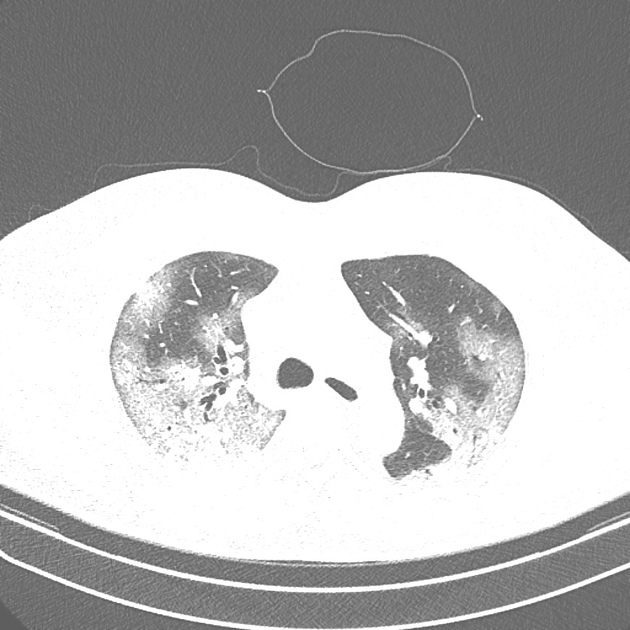
CT demonstrates bilateral, multilobar large areas of mixed consolidations and ground-glass opacities with reticular and interlobular septal thickening featuring the crazy-paving pattern. The lesions are mostly in the peripheral parts of the lungs.
This patient tested positive for SARS-CoV-2, and the CT findings are consistent with the transition from the end of the progressive stage (stage 2) to the emerging peak stage (stages 3) COVID-19 pneumonia.


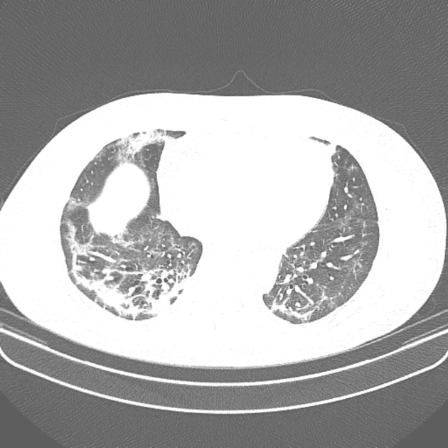
CT shows improvement in the disease course, characterised by partial regression of the bilateral, multilobar areas of airspace consolidations leaving ground-glass opacities, with some subpleural parenchymal bands already emerging.
CT demonstrates findings consistent with the end of the peak stage (stage 3) and the beginning of the late/absorption stage (stage 4) of COVID-19 pneumonia.
Case Discussion
Covid-19 is a viral infectious disease in which most patients present with respiratory symptoms 1-6. Radiologists should become familiar with the tomographic temporal stages of this disease 1-6. The most prominent feature of the progressive stage (stage 2, day 5-8 from symptoms onset) are ground-glass opacities (GGOs) with the crazy-paving pattern; In the peak stage (stage 3, day 9-13), consolidation becomes prevalent; After 14 days, (stage 4), in the late or absorption stage the lesions undergo a gradual absorption, leaving tenuous residual GGOs and subpleural parenchymal bands 1-6.
This case illustrates the temporal CT changes of progressive / peak stages (stages 2/3) with gradual improvement to the beginning of the absorption stage (stage 4) of COVID-19 pneumonia.


 Unable to process the form. Check for errors and try again.