
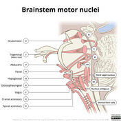


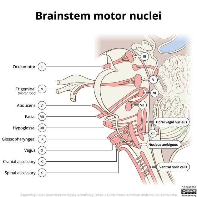
Illustration depicting the cranial nerves that contain motor fibres and the respective nuclei.
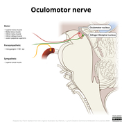


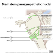
Illustration depicting the oculomotor nerve and nucleus as well as the Edinger-Westphal nucleus.


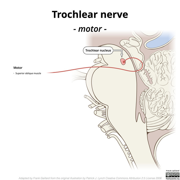
Illustration depicting the trochlear nerve and nucleus.
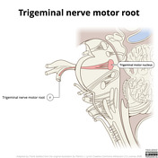


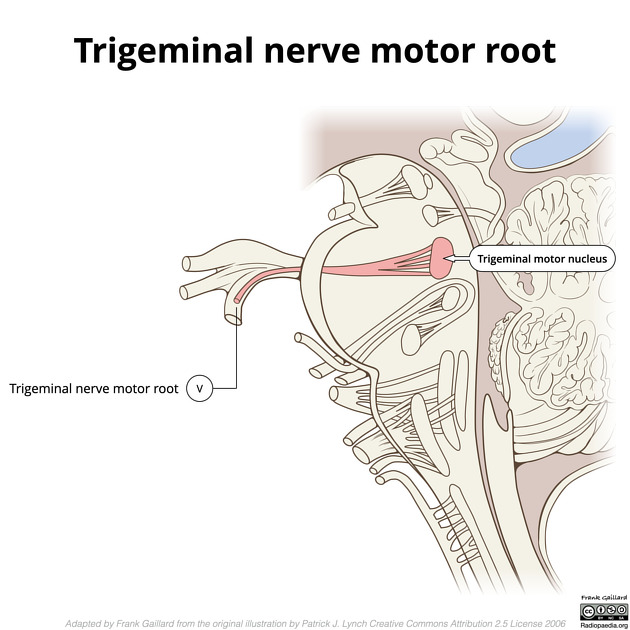
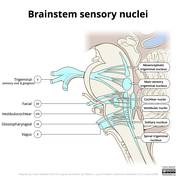
Illustration depicting the motor root of the trigeminal nerve and motor nucleus.
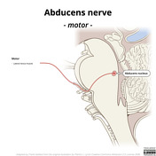

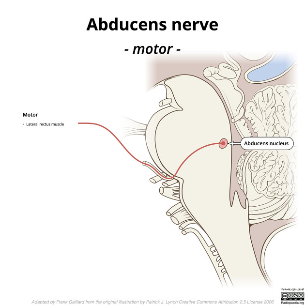
Illustration depicting the abducens nerve and nucleus.
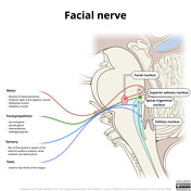
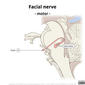


Illustration depicting the facial nerve and nucleus and nervus intermedius.

Illustration depicting the vestibulocochlear nerve and nuclei.
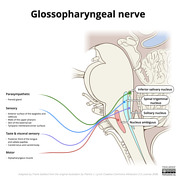

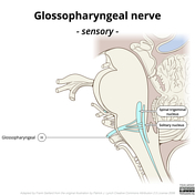
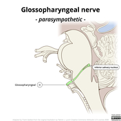

Illustration depicting the glossopharyngeal nerve and nucleus ambiguus.


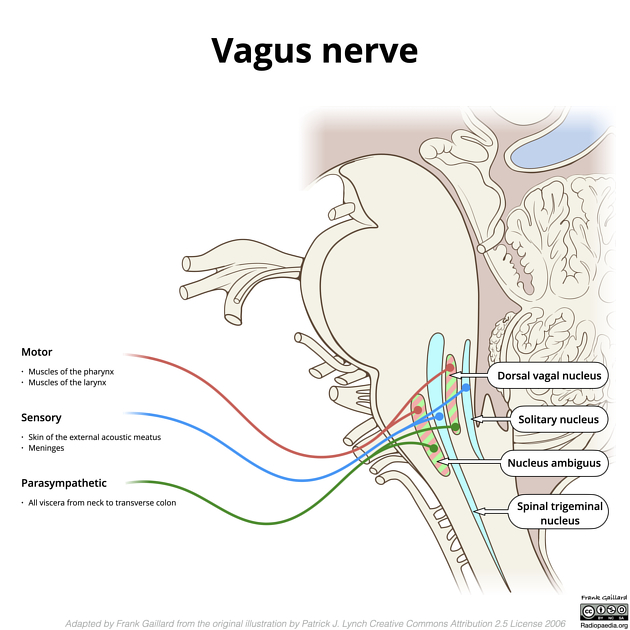
Illustration depicting the vagus nerve and nucleus ambiguus and dorsal vagal nucleus.


Illustration depicting the accessory nerve, both cranial and spinal components.


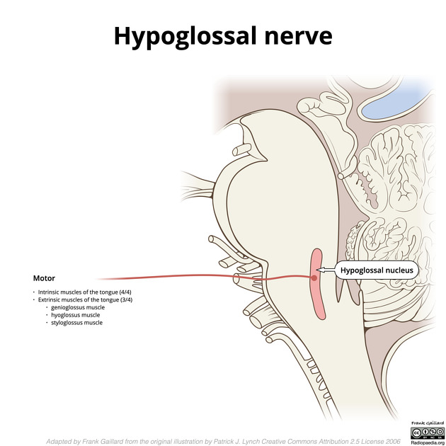
Illustration depicting the hypoglossal nerve and nucleus.
Case Discussion
These illustrations are adapted from the original illustration by Patrick Lynch released on Creative Commons Attribution 2.5 License 2006 and available on Wikimedia commons here.
Changes were made by Frank Gaillard, and include:
- repositioning of Edinger-Westphal nucleus
- adding trigeminal motor root
- adding glossopharyngeal fibres from nucleus ambiguus
- adding cranial accessory
- adding vagal fibres from dorsal vagal nucleus
- adding parasympathetic nuclei and nerves
- adding sensory nuclei and nerves


 Unable to process the form. Check for errors and try again.