
Presentation
Found unconscious on a driveway with obvious signs of head injury. GCS 5 E3V1M1.
Patient Data
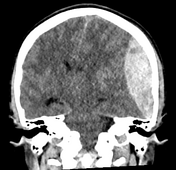

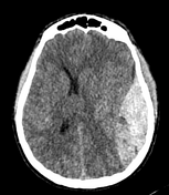
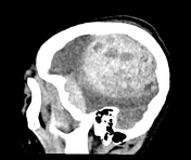

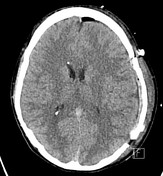
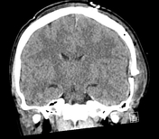
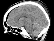
A large heterogenous convex-shaped hematoma is demonstrated in the left convexity. This is an extradural hematoma with a smaller subdural hematoma component. There is an overlying minimally displaced left parietal skull fracture. Significant midline shift of 14 mm to the right is noted.
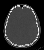
Postoperative CT head

There has been an interval left hemispheric craniotomy. This has resulted in markedly reduced hemorrhagic component previously overlying the left frontoparietal region. There is significant reduction of the rightward midline shift.
Case Discussion
This case demonstrates a large traumatic left convexity extradural hematoma with overlying minimally displaced fracture of the left parietal bone. The patient underwent emergency craniotomy surgery and the postoperative CT confirms interval evacuation of the extradural hematoma with resultant reduction in mass effect and midline shift.


 Unable to process the form. Check for errors and try again.