
Presentation
The patient was sent on MR enterography after coloscopy revealed inflammatory reactions in the walls of the caecum (in the region of the ileocaecal angle). The colonoscope did not pass into the terminal ileum. A biopsy has taken place.
Patient Data


MR enterography with oral contrast using mannitol.
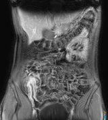
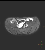
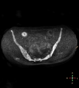
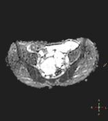
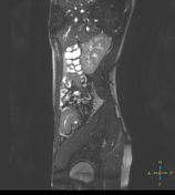
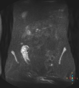
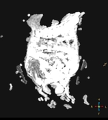
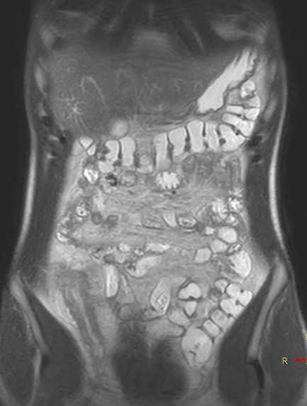
There is wall thickening (max thickness 10 mm, length of changes more than 80 mm) in the terminal ileum, ileocaecal zone and proximal caecum. In addition, the affected walls of the ileum restrict diffusion, indicating active inflammation.
The intestinal walls at this level are inflamed with reactive changes in the surrounding tissues, the mesentery, and the formation of an infiltrate in the ileocaecal angle, up to 42 mm in size. After injection of a contrast agent - layered enhancement pattern. In the same area, lymph nodes up to 6 mm in size are traced.
The appearances are highly suggestive of Crohn's disease (severe activity).
Histology: tumour not found, inflammatory changes possibly associated with Crohn's disease.
Case Discussion
Тhickening of the wall ileum up to 10 mm with restricted diffusion, inflammatory mass, layered contrast enhancement pattern, is compatible with the histology of Crohn's disease.


 Unable to process the form. Check for errors and try again.