
Presentation
Primigravida. Gestational age 14 weeks 0 days. No complaints.
Patient Data


Single live fetus with growth corresponding to the gestational age. The left side of the fetal abdomen shows two adjacent/ bilobed kidneys measuring 11 mm in length. Two separate small cystic spaces possibly correspond to the two renal pelvis.

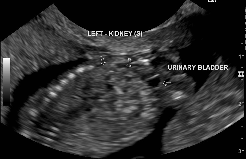
Bilobed left renal kidney(s) measuring 56 mm in length. Normal corticomedullary differentiation. No hydronephrosis or cysts. A notch is present between two renal parenchyma. Normal urinary bladder. Low-lying adrenal on the right side due to an absent kidney in the right renal fossa.
Case Discussion
A 14-week scan raised the possibility of crossed renal ectopia. 20-week scan which was done somewhere else confirmed the finding (not uploaded, no copyright). It is easy to see crossed as well as fused renal ectopia at a 37-week scan. There was no associated anomaly.


 Unable to process the form. Check for errors and try again.